
A 70 year old man with cough and fever…what do you see?

scroll down for an answer!
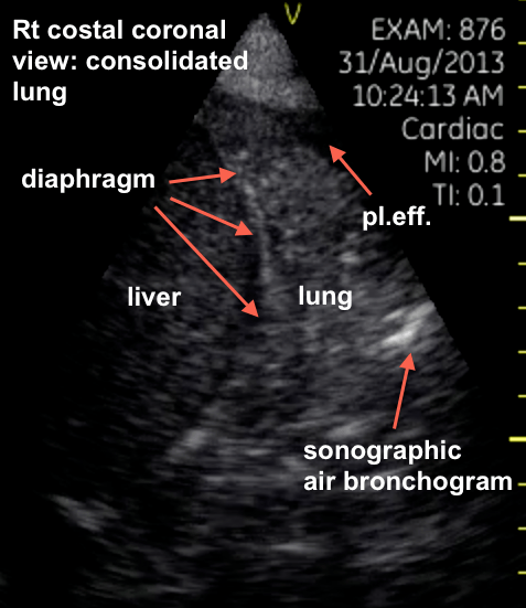
The ultrasound diagnosis is of a consolidation with a small pleural effusion. This can be referred to a “hepatization” as the appearance becomes quite similar to an unaerated organ. A sonographic air bronchogram is seen as well. Clinical correlation with the history and bloodwork strongly suggests a pneumonia.
Consolidated lung at the right basis with a small pleural effusion. The red arrow indicate something similar to an air bronchogram (althoug not very clear in a still image). From a sonographic point of view (and also clinically) pneumonia is in the differential diagnosis.
Perfect!