A VExUS Mini-Tutorial. #FOAMed, #POCUS

So a lot of people have been asking for a VExUS tutorial, and since our paper was just accepted, I figured it’s a pretty good time to do it! Thanks to Dr. Ian Ajmo of FrancoFoam fame who put on his Hollywood director’s hat and filmed it!

Here is the classification that has been validated:

I’m attaching our chapter on venous congestion below as well.


Of course, Andre Denault, William Beaubien Souligny, Rory Spiegel, Korbin Haycock and myself will be running VExUS workshops at H&R2020. There aren’t many spots left!

click here for the conference page!


Fluid Philosophy & Physiology. #STOWEEM20 Lecture! #foamed #foamcc


So I love the UVM EM Update at Stowe. It’s a great little conference, run by my good friend and all around awesome guy Peter Weimersheimer (VTEMsono) ED Pocusologist, and his super team including Kyle DeWitt (@emergpharm), Meghan Groth (ENpharmgirl) and Mark Bisanzo (@mbisanzo). It’s a smooth running show with some really amazing speakers where I always learn a bunch. Had the chance to finally meet Sergey Motov (@painfreeED) and learn from an awesome opioid lecture. And it’s always great to hang with Josh (@PulmCrit) and listen to the pearls!

So here is my fluid talk. The Keynote pdf is just below. Hope there’s a useful tidbit or two in there!


PDF: STOWE20 Fluids

So thanks again to Peter and his crew! And the Stowe attendees, as we had mentioned, get to attend H&R2020 at the in-house rate! (Please choose the Special Members rate)


Cheers!

Philippe


VEXUS Lite: Screening for Venous Congestion with Handheld POCUS. #FOAMed, #FOAMus

So recently a colleague asked me about one of my twitter posts where I had put a clip of doing venous congestion assessment using a handheld – which is without pulsed Doppler (PW).  Since VEXUS is predominantly based on Doppler findings, seems like 2D and colour might not cut it, but can it be done in a screening or “lite” fashion?

Definitely. Here is a mini-discussion about it, and some clips below to illustrate.


Pulsatile PV

Clearly Pulsatile PV likely near 100%

Ascites, plethoric IVC, pulsatile PV, markedly abnormal HV with “police siren” appearance due to substantial retrograde flow – likely VExUS 3 or C.

Normal looking HV

Markedly abnormal HV


Love to hear some questions or comments!


of course, lots of VExUS discussions with William Beaubien Souligny, Andre Denault, Rory Spiegel, Korbin Haycock and myself at H&R2020!

cheers


Philippe


The Resus Tracks: Trans-Pulmonary Dilution Catheters in the ED…myth or reality? #FOAMed, #FOAMer

So anyone who knows Korbin (@khaycock2) realizes he is a true trailblazer in the ED, essentially doing cutting edge critical care from the get go in his shock patients. In my mind this should be the goal for any critically ill patients, that they get the highest level care right at entry and for however long they may be staying in the ED until they get to the ICU.

So today, I was really happy to corner Korbin lounging somewhere in sunny California (as 6 inches of snow come down hard in Montreal) to tell me how he is using this technology in his resus patients.


So this has got me interested in using this technology. I see it as an early warning signal that your patient may be less fluid tolerant than you may think, and that the signs of pulmonary fluid intolerance I use (oxygen requirement, appearance of B lines (FALLS Protocol-style), etc…) have yet to manifest.

So I’m looking forward to hearing Korbin explain this further (during H&R2020!) and in actual cases where the change in management is clear.


cheers


Philippe


H&R2019 Lecture Series: A Christmas Special TripleHeader! #FOAMed, #FOAMer

Happy holidays to all, here is a little gift from H&R2019!

So here’s a gem from Kylie, who is my reference point for GI POCUS. Ever since this lecture I’ve been working on upping my abdominal scanning capabilities!

GI POCUS with Kylie Baker


Also, if you’ve been working on your Doppler applications, best do so while truly understanding it, both strengths and pitfalls. Here’s the physiology master shedding some light and some insights.

Advanced Doppler with Jon-Emile Kenny


And finally, for the airway fans, here’s Rory Spiegel (@EMNerd) sharing his skills with the bougie.

The Art of the Bougie with Rory Spiegel


cheers

Philippe


For more lectures that will change the way you practice medicine, do try to make it to H&R2020! It is the very ethos of this small conference, to exchange with docs pushing the envelope of acute care as well as like-minded peers. This isn’t the conference to go to if you want to see what most people do and review guidelines. This is the one to go to if you want to be on the cutting edge. Only 100 spots, has sold out every year.  For more info and registration click here!

#FOAMresus Case from Amand Thind (@Thind888)

So #MedTwitter is truly an incredible forum for case discussion, where you get to exchange with literally some of the best medical minds on the planet who often also happen to be front-line clinicians in the nitty-gritty therapeutic decision-making. Here’s a discussion which I think was great. Recently, Dr. Thind has been generating some great cases and hemodynamic discussions. I thought this one was worth highlighting!
Dr Thind is an internist and currently Critical Care Hospitalist (and upcoming ICU fellow) at the Cleveland Clinic, and tweets out some great #FOAM from @Thind888 on twitter.
Case:
OK, let’s give this a shot. Here’s a ‘hemodynamics special’. Saw this case a couple weeks ago. A lot of decision making was based on educated guesses so it should be a good one for discussion. – 51 yo woman being worked up on the floor for chronic diarrhea, moved to ICU for hypoxia.
Dyspnea progressed over few hours. Vitals significant for tachycardia (140s) and hypotension (MAP in low 60s). On arrival, SBP 60s – improved with fluid bolus. CXR attached. Patient has H/O of pericardial effusion for several months that has been managed conservatively. 
The patient has an official ECHO performed on arrival in ICU (images attached). IVC difficult to assess but about 2cm without collapse. Lung US – diffuse B lines. 
OK so right there a flag goes up for me. A plethoric IVC means something is wrong. Sounds too vague maybe, but you need to find the reason for this, as it likely has therapeutic implications. Let’s see what comes up.
Modifed A5C.
LVOT doppler

Image

Image

Image

CXR

Image

Pressing questions –
(i) Is it hydrostatic or increased permeability pulmonary edema?
(ii) Fluids, diuresis, or none?
(iii) Would CPAP help?
(iv) Drain the pericardial effusion?
(v) What about that LVOT doppler? 
Mitral inflow velocities and TDI attached. M-mode through PLAX almost uninterpretable. Lung infiltrates are new so less likely lymphangitic carninomatosis. Note: ScVo2 = 40s. Another Q to ponder on –
(vi) Is tamponade typically associated with hydrostatic pulmonary edema?

Image

Image

Image

Image

Perhaps this slowed up (0.5x) A3C loop will help with that LVOT doppler!

Great discussion as expected. Lets discuss:
Q4. Is it tamponade? – This is not a slam dunk. Chamber collapse can sometimes be controversial. In these situations I try my best to get MV E-wave variation. I think our tech got a decent signal. But note these are fused E/A waves.
The first thing I look at to screen for tamponade is the IVC. Tamponade is an obstructive form of shock, dependant on the intrapericardial pressure exceeding the right atrial pressure. If it does, unless respiratory efforts are extreme, the IVC should become plethoric. Hence, the absence of such would make the effusion – given the current RA pressure – NOT tamponade. Yet again, another point scored by the IVC for usefulness.
Although I don’t see why we can’t use fused waves for this purpose (couldn’t find anything on it in the literature). Note that in spite of the cardiac motion, the mitral inflow variation is <25% (~23%). It’s close though, and certainly seems to have increased from 3 days ago.

Image

The cardiologist (understandably) was non-committal and read it as “possible early tamponade”.
Q5. What about LVOT doppler? A good M-mode could not be obtained but the A3C in 6/ shows SAM. The report mentioned “chordal SAM” but I think you can clearly see “valvular SAM” too.
Chordal SAM is SAM of the chordal apparatus (you could see it bumping against the septum in 6/). It is (typically) NOT hemodynamically significant (PMID: 27241937). – When we see mitral SAM, it is important to quantify its hemodynamic effects – with LVOT peak gradient via CW.
In HOCM, DLVOTO is defined by an LVOT gradient of >30; >50 is considered severe. Our patient had a gradient of ~70. Although classically a/w HCM, SAM can be seen in anyone with thick, hypercontractile, underfilled LV. Tachycardia further hampers LV filling (PMID: 27726435).
Mitral SAM is often a/w MR – this acute MR can cause flash pulmonary edema. These patients may actually need fluids (to help with SAM) to fix there hydrostatic pulmonary edema!! (PMID: 20661209). However, our patient only had trace MR (you could see it in 1-2 CD frames).
Working theory (similar to Lars) – Chronic stable pericardial effusion –> diarrhea (pt had 15 BMs the day before the admission) –> reduced venous return –> brought the patient at the verge of low-pressure tamponade (PMID: 16923755) –> further reduction in LV filling  —> reduced stroke volume –> adrenergic drive causing tachycardia and increased inotropy –> all factors culminating in mitral SAM and DLVOTO.
This also explains the low ScVO2. Note – CPAP would further reduce venous return (Q3) so wouldn’t help, may hurt.
Now the most important Qs: why pulmonary edema and what to do about it (Q1 and 2). As tamponade causes impedance to venous return, it is not typically associated with high LAP and hydrostatic pulmonary edema (Q6).
But first, let’s check out another CW tracing. Any thoughts?

Image

Image

This is a CW beam through LV apex and mitral valve – typically performed to assess mitral inflow and MR velocities and is part of the standard ECHO exam. However, the tracing is not typical for MR (late peaking, dagger shape). Remember, CW does not have depth resolution.

Image

This is likely mid-cavitay/intra-ventricular obstruction. This is caused by complete mid-systolic obliteration of LV cavity (see PSAX) causing obstruction to the apical systolic flow. Again, seen in hypercontractile, underfilled, thick LV – e.g. sepsis (PMID: 26082197).
Finally – what does the ECHO tell us about LV filling pressures? – E/A ratio: As Lars pointed out, an E/A < 0.8 usually means normal LAP. However, the exception to this is sinus tach. This was shown in a study by none other than Dr. Nagueh (PMID: 9778330). (Also, see image)

Image

The idea is that when early filling (E) is incomplete due to short diastolic time, the LA remains “full” at the time of the atrial kick – causing higher A velocities. NB: In that paper, E/E’ > 10 had a specificity of 95% for elevated LAP in ST. In our case: E/E’ = 75/5 = 15!

Image

Potential contributors of high LAP – (i) SAM-associated MR – ‘trace’ in this ECHO but maybe we didn’t catch it. (ii) Tachycardia – E’ is 5 suggestive of delayed relaxation. Tachycardia causes “incomplete relaxation”. (iii) High afterload – high-grade dynamic obstructions.

Image

So at this point, it’s still contentious but I have my money on hydrostatic pulmonary edema. Will detail our interventions and the remaining course in a bit. …Sorry to make this long but I think it’s worth it!
Now for the home stretch, the remaining course: We realized pericardiocentesis may be required soon but wanted to see if volume helps with (i) Peri-tamponade (ii) Dynamic obstructions. It helped a little – O2 requirements went from 60% HF to 6L NC. BP okay but still tachy.
Day 2: We pushed 2.5 mg metop x2 with concurrent ECHO. LVOT gradient improved from 70s to ~10! (I did not compare mid-cavitary gradient, apologies). Started on 25 bid of PO metop later that night. HR now 90s Day 3: Official ECHO shows improved but persistent gradients.

Image

Image

Evaluation of tamponade was similar to previous ECHO but E-wave velocity variation now 38% –> elective pericardiocentesis: 550 cc removed. Fluid was transudate We also tapped a small pleural effusion pocket: transudate, cx negative (again goes with hydrostatic pulmonary edema).

Image

Day 3 (contd): inc metop to 50 Q12H to blunt the gradients.
Day 4 – HR in 80s. ECHO shows no DLVOTO and non-significant mid-cavitary gradient. Oxygenation improved but still not normal. Why?! Check the E-velocity post-pericardiocentesis: it has jumped to 120 with E/A > 1.

Image

So why is the LAP still high despite no significant dynamic obstruction? – Patients with chronic pericardial effusion may have chronically impaired diastolic filling –> low output –> volume retention (basic CHF physiology). When pericardial restraint suddenly released ––> increased LV preload –> high LAP.
Originally discussed elegantly here: PMID 6877287.
This is especially true if the LV has some baseline dysfunction. Day 5 – We started diuresis! The obvious risk was to precipitate the dynamic obstructions –> metop increased to 50 Q8H.
Day 7: Excellent diuresis (~2-3L negative per day). Hemodynamics stable (SvCO2 normal). Resting HR 60s – 70s. Follow-up ECHO confirmed no dynamic obstructions (see image). Day 8: Finally on room air. Pulmonary infiltrates improved (image). All cx remained negative.

Image

Image

Some dogmalysis offered by this case – – Fluids (probably) helped the pulmonary edema; CPAP/diuresis may have worsened. – IV metop contraindicated in hypotension? Not in this case – Sometimes you may have to diurese someone who recently had DLVOTO, as discussed above.
This case highlights the cognitive flexibility required to deal with hemodynamic puzzles. One thing I would’ve done different is be more aggressive with metop early on as it made a huge difference with DLVOTO. This was quite a ride. Hope you had fun. Feel free to share! 
Much kudos to the treating team, I think this was excellently managed. As Amand says, cognitive flexibility ias absolutely key in assessing hemodynamics, particularly in the grey zones when multiple processes occur and co-exist. Managing this type of case using a recipe-based approach and without POCUS could have let to a poor outcome. 
Now the POCUS used in this case is on another level. Very impressive and allowing incredible insight and certainly many potentially clinically useful Doppler analysis tips for LVOTO and LAP assessment. 
In the end, I think that there were three pathologies, (a) tamponade physiology, (b) dynamic LVOTO, exacerbated by (c) hypovolemia (diarrhea)  I might have approached this differently, had I seen a truly plethoric IVC. In such a case, one can easily see how tamponade physiology would contribute to LVOTO in two ways by creating intracardiac hypovolemia, hence worsening LVOTO both by decreasing LV preload and by the compensatory tachycardia. My first approach would probably have been to drain the pericardial effusion, and reassessing the hemodynamics afterwards, but correcting the intravascular deficit was necessary.
The other important thing this case re-emphasize is that tamponade is not a static diagnosis but a physiological spectrum. For the same given effusion (read intrapericardial pressure – IPP), it is the RA pressure that will determine whether overt tamponade develops. In this patient, it is very likely that a day earlier, there was no frank tamponade, but that after some diarrheal volume loss, the RAP dropped, and now IPP > RAP.  It is important to know this because if you have an effusion and a fairly full IVC, one needs to be very careful with anything that can drop the RAP, meaning diuretics and vasodilators, because these can easily turn pre-tamponade into overt shock.  And, as this case illustrates so well, you might even end up with LVOTO and pulmonary edema!  Which is one of the myriad reasons one should have a basic POCUS exam in every acutely ill patient. These are things a resucitationist needs to know and prepare for.
cheers and thanks again to Dr. Thind!
Philippe

H&R2019 Lecture Series: Denault on POCUS and Delirium! #FOAMed, #FOAMcc

So the pundits still try to claim the lack of evidence for the use of POCUS, bla, bla bla. Just wait till they get a load of this: POCUS in delirium? Master Andre Denault introduces us here to a completely new way of assessing a little known potential aetiology of acute delirium.


Here it is, certainly one of the most interesting and forward-thinking lectures of H&R2019:

Denault on POCUS and Delirium.


Don’t forget The Hospitalist & The Resuscitationist H&R2020 is happening May 20-22 and registration is open! Seating is limited…

cheers!

Philippe

A few words on venous congestion, thresholds, and physiology. #FOAMed


So I’m really glad to see that recently, a lot of discussion has been taken place on the topic of right sided failure and venous congestion, which has huge clinical applications. Even more so, the fact that a lot of individual practitioners have taken this on and have been applying it clinically with physiological results is really amazing.

So a common question that has been popping up revolves around clinical thresholds of significance, and I thought it was worth clarifying that we need to stay away from a pure threshold approach, but rather try to embrace a holistic cardio pulmonary and whole body assessment.

So here’s my two cents:

Thank you, love to hear any comments!

Philippe

ps obviously, this type of discussion will be what H&R2020 will be chock full of!


H&R2019 Lecture Series: Weimersheimer (@VTEMsono) on Massive Transfusion. #FOAMed, #FOAMer

Sorry for the delay, been bogged down in getting H&R2020 off the ground! But here is another goodie from H&R2019, my good friend Peter W. on an ever-important trauma topic.  Enjoy!

Weimersheimer on MT – H&R2019.


cheers


Philippe

ps for anyone interested, H&R2019 On Demand can be found here!

POCUS Skill: Bedside Percutaneous Cholecystostomy. #FOAMed, #FOAMcc

So one thing we all pretty much agree on is the importance of source control. Biliary sepsis is one of the more common causes of intra-abdominal sepsis, and among those, there is a not insignificant proportion of cases where a percutaneous drainage procedure is indicated, often related to an elevated surgical risk.

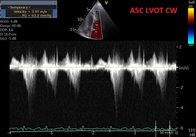
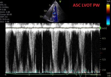
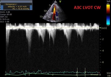
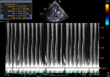
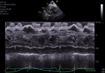
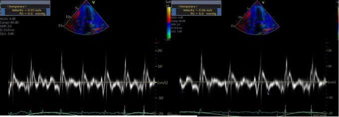
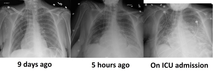
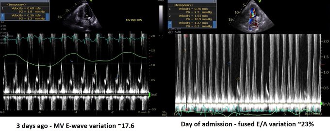
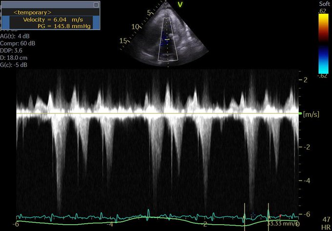
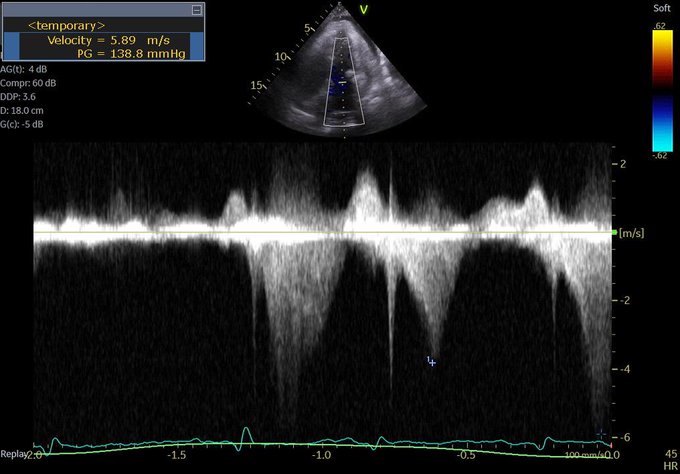
This is the case of a 90 year old man with severe aortic stenosis and a perforated cholecystitis with sepsis (AKI, delirium, coagulopathy) admitted to our ICU. Due to the aortic stenosis, surgical mortality was felt to be quite elevated, hence a percutaneous procedure was done.

I am sharing this to make the case that a percutaneous cholecystostomy is not outside the reasonable skill set of a clinician who is both POCUS competent and has solid guided procedural experience (central lines, thoracic or abdominal pigtails, etc) and in my opinion falls into that same category as pericardiocentesis. All the more so for clinicians working in community hospitals without the luxury of a 24/7 IR team, because in many cases, it is simply not reasonable to wait many hours for source control – the fact that the patient may make it alive to the next morning to have a drainage procedure is not relevant, as the ongoing sepsis over several hours may be something he or she does not always recover from in the ensuing days and is not a risk worth taking unless there is no other viable option. In our center the critical care physicians perform this intervention when IR is not available.

Here, an in-plane approach was chosen with a trans-hepatic route in order to avoid potential peritoneal spillage.

POCUS Pearls: 

(1) Always visualize the guidewire inside the intended space.

(2)When dilating, make sure the proximal part of the guidewire within the target area “disappears” ultrasonographically, confirming entry of the dilator. Why? In some cases the wall may give more resistance (particularly an inflammed pericardium) and the dilator may remain outside – cannulation with the catheter will be impossible.

Procedure:


POCUS Clips


And the nasty stuff:


Some relevant articles:

https://www.ncbi.nlm.nih.gov/pubmed/12040818

https://www.ncbi.nlm.nih.gov/pubmed/29519331


Love to hear of others’ experience,


cheers

PS if anyone wants a perc chole workshop at H&R2020 , let me know!


Philippe