HR24: Acute Inpatient Medicine – Next Level

June 15, 2024, 10-4 EST – Online Webinar Only

Due to the tremendous amount of work required to put together a large live event, we’ve decided to skip 2024 in terms of a full-fledged H&R, but HR23’s Inpatient Medicine section, put together by none else than Katie Wiskar, was a real firecracker, and we felt that the spark to bring high level bedside physiology to the clinician on the wards should not be allowed to fizzle. So, while waiting for HR25 (May 2025), Katie once again worked her magic to put together a one-day webinar that packs a academic and clinical punch.

Preliminary Schedule:

H&R 2024 Outline: Acute Inpatient Medicine – Next Level! 

Date: June 15 10am-4:30pm EST (7am-1:30pm PST) 

Format: 

  • one-day, 6.5 hours, online only
  • Pre-recorded lectures
  • Live Q&A

The Day

  • 10am-10:15am: Intro and welcome (15min)
  • Track 1 (90min) – POCUS et al
    • 10:15am-10:35am: POCUS for shock – Ross Prager
    • 10:35am-10:55am: VEXUS tips and tricks – Abhilash Koratala
    • 10:55am-11:15am: Ventilators 101 – Segun Olusanya
    • 11:15am-11:35am: Q&A 
  • Track 2 (90 min) – Fluids and physiology
    • 11:35am-11:55am: Dysnatremias in cirrhosis – Eduardo Argaiz
    • 11:55am-12:15pm: Top 10 diuretic mistakes – Frederik Verbrugge 
    • 12:15pm-12:35pm: Physiology of the shock patient – Ashley Miller 
    • 12:35-12:55pm: Q&A
  • 12:55pm-1:30pm: Break (35min)
  • Track 3 – The decompensating ward patient
    • 1:30pm-1:50pm: The sick aortic stenosis patient – Trina Augustin
    • 1:50pm-2:10pm: The sick pulmonary HTN patient – Matt Siuba
    • 2:10pm-2:30pm: The seizing ward patient – Casey Albin
    • 2:30pm-2:50pm: Q&A
  • Track 4 – Practical pearls
    • 2:50pm-3:10pm:  Palliative Medicine pointers – Brittany Rance
    • 3:10pm-3:30pm:  Top 5 Geriatric Pearls on the ward – Gurmeet Sohi
    • 3:30pm-3:50pm: Sepsis: what we should be doing – Sara Crager
    • 3:50pm-4:10pm: Q&A
  • 4:10-4:30pm: Concluding remarks (20min)

Also some online-only BONUS lectures – How-To VExUS by Dr. Taweevat Assavapokee!

Registration is OPEN! https://ccusinstitute.wixsite.com/ccus/events/hr2024-acute-inpatient-medicine-next-level-online-webinar

Cheers!

The Resus Tracks: Trans-Pulmonary Dilution Catheters in the ED…myth or reality? #FOAMed, #FOAMer

So anyone who knows Korbin (@khaycock2) realizes he is a true trailblazer in the ED, essentially doing cutting edge critical care from the get go in his shock patients. In my mind this should be the goal for any critically ill patients, that they get the highest level care right at entry and for however long they may be staying in the ED until they get to the ICU.

So today, I was really happy to corner Korbin lounging somewhere in sunny California (as 6 inches of snow come down hard in Montreal) to tell me how he is using this technology in his resus patients.


So this has got me interested in using this technology. I see it as an early warning signal that your patient may be less fluid tolerant than you may think, and that the signs of pulmonary fluid intolerance I use (oxygen requirement, appearance of B lines (FALLS Protocol-style), etc…) have yet to manifest.

So I’m looking forward to hearing Korbin explain this further (during H&R2020!) and in actual cases where the change in management is clear.


cheers


Philippe


H&R2019 Lecture Series: A Christmas Special TripleHeader! #FOAMed, #FOAMer

Happy holidays to all, here is a little gift from H&R2019!

So here’s a gem from Kylie, who is my reference point for GI POCUS. Ever since this lecture I’ve been working on upping my abdominal scanning capabilities!

GI POCUS with Kylie Baker


Also, if you’ve been working on your Doppler applications, best do so while truly understanding it, both strengths and pitfalls. Here’s the physiology master shedding some light and some insights.

Advanced Doppler with Jon-Emile Kenny


And finally, for the airway fans, here’s Rory Spiegel (@EMNerd) sharing his skills with the bougie.

The Art of the Bougie with Rory Spiegel


cheers

Philippe


For more lectures that will change the way you practice medicine, do try to make it to H&R2020! It is the very ethos of this small conference, to exchange with docs pushing the envelope of acute care as well as like-minded peers. This isn’t the conference to go to if you want to see what most people do and review guidelines. This is the one to go to if you want to be on the cutting edge. Only 100 spots, has sold out every year.  For more info and registration click here!

#FOAMresus Case from Amand Thind (@Thind888)

So #MedTwitter is truly an incredible forum for case discussion, where you get to exchange with literally some of the best medical minds on the planet who often also happen to be front-line clinicians in the nitty-gritty therapeutic decision-making. Here’s a discussion which I think was great. Recently, Dr. Thind has been generating some great cases and hemodynamic discussions. I thought this one was worth highlighting!
Dr Thind is an internist and currently Critical Care Hospitalist (and upcoming ICU fellow) at the Cleveland Clinic, and tweets out some great #FOAM from @Thind888 on twitter.
Case:
OK, let’s give this a shot. Here’s a ‘hemodynamics special’. Saw this case a couple weeks ago. A lot of decision making was based on educated guesses so it should be a good one for discussion. – 51 yo woman being worked up on the floor for chronic diarrhea, moved to ICU for hypoxia.
Dyspnea progressed over few hours. Vitals significant for tachycardia (140s) and hypotension (MAP in low 60s). On arrival, SBP 60s – improved with fluid bolus. CXR attached. Patient has H/O of pericardial effusion for several months that has been managed conservatively. 
The patient has an official ECHO performed on arrival in ICU (images attached). IVC difficult to assess but about 2cm without collapse. Lung US – diffuse B lines. 
OK so right there a flag goes up for me. A plethoric IVC means something is wrong. Sounds too vague maybe, but you need to find the reason for this, as it likely has therapeutic implications. Let’s see what comes up.
Modifed A5C.
LVOT doppler

Image

Image

Image

CXR

Image

Pressing questions –
(i) Is it hydrostatic or increased permeability pulmonary edema?
(ii) Fluids, diuresis, or none?
(iii) Would CPAP help?
(iv) Drain the pericardial effusion?
(v) What about that LVOT doppler? 
Mitral inflow velocities and TDI attached. M-mode through PLAX almost uninterpretable. Lung infiltrates are new so less likely lymphangitic carninomatosis. Note: ScVo2 = 40s. Another Q to ponder on –
(vi) Is tamponade typically associated with hydrostatic pulmonary edema?

Image

Image

Image

Image

Perhaps this slowed up (0.5x) A3C loop will help with that LVOT doppler!

Great discussion as expected. Lets discuss:
Q4. Is it tamponade? – This is not a slam dunk. Chamber collapse can sometimes be controversial. In these situations I try my best to get MV E-wave variation. I think our tech got a decent signal. But note these are fused E/A waves.
The first thing I look at to screen for tamponade is the IVC. Tamponade is an obstructive form of shock, dependant on the intrapericardial pressure exceeding the right atrial pressure. If it does, unless respiratory efforts are extreme, the IVC should become plethoric. Hence, the absence of such would make the effusion – given the current RA pressure – NOT tamponade. Yet again, another point scored by the IVC for usefulness.
Although I don’t see why we can’t use fused waves for this purpose (couldn’t find anything on it in the literature). Note that in spite of the cardiac motion, the mitral inflow variation is <25% (~23%). It’s close though, and certainly seems to have increased from 3 days ago.

Image

The cardiologist (understandably) was non-committal and read it as “possible early tamponade”.
Q5. What about LVOT doppler? A good M-mode could not be obtained but the A3C in 6/ shows SAM. The report mentioned “chordal SAM” but I think you can clearly see “valvular SAM” too.
Chordal SAM is SAM of the chordal apparatus (you could see it bumping against the septum in 6/). It is (typically) NOT hemodynamically significant (PMID: 27241937). – When we see mitral SAM, it is important to quantify its hemodynamic effects – with LVOT peak gradient via CW.
In HOCM, DLVOTO is defined by an LVOT gradient of >30; >50 is considered severe. Our patient had a gradient of ~70. Although classically a/w HCM, SAM can be seen in anyone with thick, hypercontractile, underfilled LV. Tachycardia further hampers LV filling (PMID: 27726435).
Mitral SAM is often a/w MR – this acute MR can cause flash pulmonary edema. These patients may actually need fluids (to help with SAM) to fix there hydrostatic pulmonary edema!! (PMID: 20661209). However, our patient only had trace MR (you could see it in 1-2 CD frames).
Working theory (similar to Lars) – Chronic stable pericardial effusion –> diarrhea (pt had 15 BMs the day before the admission) –> reduced venous return –> brought the patient at the verge of low-pressure tamponade (PMID: 16923755) –> further reduction in LV filling  —> reduced stroke volume –> adrenergic drive causing tachycardia and increased inotropy –> all factors culminating in mitral SAM and DLVOTO.
This also explains the low ScVO2. Note – CPAP would further reduce venous return (Q3) so wouldn’t help, may hurt.
Now the most important Qs: why pulmonary edema and what to do about it (Q1 and 2). As tamponade causes impedance to venous return, it is not typically associated with high LAP and hydrostatic pulmonary edema (Q6).
But first, let’s check out another CW tracing. Any thoughts?

Image

Image

This is a CW beam through LV apex and mitral valve – typically performed to assess mitral inflow and MR velocities and is part of the standard ECHO exam. However, the tracing is not typical for MR (late peaking, dagger shape). Remember, CW does not have depth resolution.

Image

This is likely mid-cavitay/intra-ventricular obstruction. This is caused by complete mid-systolic obliteration of LV cavity (see PSAX) causing obstruction to the apical systolic flow. Again, seen in hypercontractile, underfilled, thick LV – e.g. sepsis (PMID: 26082197).
Finally – what does the ECHO tell us about LV filling pressures? – E/A ratio: As Lars pointed out, an E/A < 0.8 usually means normal LAP. However, the exception to this is sinus tach. This was shown in a study by none other than Dr. Nagueh (PMID: 9778330). (Also, see image)

Image

The idea is that when early filling (E) is incomplete due to short diastolic time, the LA remains “full” at the time of the atrial kick – causing higher A velocities. NB: In that paper, E/E’ > 10 had a specificity of 95% for elevated LAP in ST. In our case: E/E’ = 75/5 = 15!

Image

Potential contributors of high LAP – (i) SAM-associated MR – ‘trace’ in this ECHO but maybe we didn’t catch it. (ii) Tachycardia – E’ is 5 suggestive of delayed relaxation. Tachycardia causes “incomplete relaxation”. (iii) High afterload – high-grade dynamic obstructions.

Image

So at this point, it’s still contentious but I have my money on hydrostatic pulmonary edema. Will detail our interventions and the remaining course in a bit. …Sorry to make this long but I think it’s worth it!
Now for the home stretch, the remaining course: We realized pericardiocentesis may be required soon but wanted to see if volume helps with (i) Peri-tamponade (ii) Dynamic obstructions. It helped a little – O2 requirements went from 60% HF to 6L NC. BP okay but still tachy.
Day 2: We pushed 2.5 mg metop x2 with concurrent ECHO. LVOT gradient improved from 70s to ~10! (I did not compare mid-cavitary gradient, apologies). Started on 25 bid of PO metop later that night. HR now 90s Day 3: Official ECHO shows improved but persistent gradients.

Image

Image

Evaluation of tamponade was similar to previous ECHO but E-wave velocity variation now 38% –> elective pericardiocentesis: 550 cc removed. Fluid was transudate We also tapped a small pleural effusion pocket: transudate, cx negative (again goes with hydrostatic pulmonary edema).

Image

Day 3 (contd): inc metop to 50 Q12H to blunt the gradients.
Day 4 – HR in 80s. ECHO shows no DLVOTO and non-significant mid-cavitary gradient. Oxygenation improved but still not normal. Why?! Check the E-velocity post-pericardiocentesis: it has jumped to 120 with E/A > 1.

Image

So why is the LAP still high despite no significant dynamic obstruction? – Patients with chronic pericardial effusion may have chronically impaired diastolic filling –> low output –> volume retention (basic CHF physiology). When pericardial restraint suddenly released ––> increased LV preload –> high LAP.
Originally discussed elegantly here: PMID 6877287.
This is especially true if the LV has some baseline dysfunction. Day 5 – We started diuresis! The obvious risk was to precipitate the dynamic obstructions –> metop increased to 50 Q8H.
Day 7: Excellent diuresis (~2-3L negative per day). Hemodynamics stable (SvCO2 normal). Resting HR 60s – 70s. Follow-up ECHO confirmed no dynamic obstructions (see image). Day 8: Finally on room air. Pulmonary infiltrates improved (image). All cx remained negative.

Image

Image

Some dogmalysis offered by this case – – Fluids (probably) helped the pulmonary edema; CPAP/diuresis may have worsened. – IV metop contraindicated in hypotension? Not in this case – Sometimes you may have to diurese someone who recently had DLVOTO, as discussed above.
This case highlights the cognitive flexibility required to deal with hemodynamic puzzles. One thing I would’ve done different is be more aggressive with metop early on as it made a huge difference with DLVOTO. This was quite a ride. Hope you had fun. Feel free to share! 
Much kudos to the treating team, I think this was excellently managed. As Amand says, cognitive flexibility ias absolutely key in assessing hemodynamics, particularly in the grey zones when multiple processes occur and co-exist. Managing this type of case using a recipe-based approach and without POCUS could have let to a poor outcome. 
Now the POCUS used in this case is on another level. Very impressive and allowing incredible insight and certainly many potentially clinically useful Doppler analysis tips for LVOTO and LAP assessment. 
In the end, I think that there were three pathologies, (a) tamponade physiology, (b) dynamic LVOTO, exacerbated by (c) hypovolemia (diarrhea)  I might have approached this differently, had I seen a truly plethoric IVC. In such a case, one can easily see how tamponade physiology would contribute to LVOTO in two ways by creating intracardiac hypovolemia, hence worsening LVOTO both by decreasing LV preload and by the compensatory tachycardia. My first approach would probably have been to drain the pericardial effusion, and reassessing the hemodynamics afterwards, but correcting the intravascular deficit was necessary.
The other important thing this case re-emphasize is that tamponade is not a static diagnosis but a physiological spectrum. For the same given effusion (read intrapericardial pressure – IPP), it is the RA pressure that will determine whether overt tamponade develops. In this patient, it is very likely that a day earlier, there was no frank tamponade, but that after some diarrheal volume loss, the RAP dropped, and now IPP > RAP.  It is important to know this because if you have an effusion and a fairly full IVC, one needs to be very careful with anything that can drop the RAP, meaning diuretics and vasodilators, because these can easily turn pre-tamponade into overt shock.  And, as this case illustrates so well, you might even end up with LVOTO and pulmonary edema!  Which is one of the myriad reasons one should have a basic POCUS exam in every acutely ill patient. These are things a resucitationist needs to know and prepare for.
cheers and thanks again to Dr. Thind!
Philippe

H&R2020: The Hospitalist Course. #Hresus20

As with all continuing medical education events, this one will make you a better physician. But we know who you are: It’s 2am. Your pager is blowing up. You go to the floor with 3 simultaneously crashing patients. We’ve all been there. As a result, we’ve collectively designed this educational event to bring together an ultimate think-tank on how to improve your clinical management of all those things that make us scared at night, and even in the day… If it’s an organ that can fail, we’ve got you covered for a solid, easy, memorable approach to how to support it.

Mixed throughout the day will be cases to solidify your newly acquired clinical pearls & hands on stations with all the toys you need to stabilize your patients.

Hosted by The IBCC (Internet Book of Critical Care) co-creators Adam Thomas and Josh Farkas – also the man behind PulmCrit – this is going to be jam-packed with physiology, clinical pearls, interactive and case-based. If you take care of patients on the wards, this is one not to miss.

Co-directors: Adam Thomas & Josh Farkas.

PRELIMINARY PROGRAMME

Hypotension on the wards – Adam Thomas reviews the physiology of shock, the use of invasive & non-invasive monitoring, POCUS & “how your radiologist can help”, initial stabilization (hint, it is not just giving fluid), the hunt for & elimination of hypovolemic, cardiogenic, obstructive shock & distributive shock, as well reviewing the role of hormones and regulatory cytokines as well as how this can potentially be modulated.

Recognizing Illness at a Glance – in this interesting talk, Daniel Kaud shares his data-linkage and pattern recognition skills on common but important pathologies, to help clinicians develop rapid muscle memory and make elusive diagnoses.

Initial stabilization of respiratory failure – this can be one of the most harrowing and time-critical clinical scenarios facing the hospitalist, before the critical care team can take over or the patient can be transferred. Adam Thomas takes participants thru the identification of respiratory failure, the rule of 2s in type & treatment of respiratory failure and the right tools for the job on the wards.

Managing the Congestive Heart Failure Patient – here, Philippe Rola introduces a physiologic and POCUS-based approach to the management of the admitted CHF patient, particularly with the management of effusions and venous congestion.

Physiologic approach to Renal Failure – nephro-intensivist Sharad Patel drags the management of this common disease into this century and will share a rapid approach based on evidence, physiology and the efficiency that POCUS brings to bedside diagnosis and clinical decision-making.

The Biliary patient – whether neoplasia or lithiasis, these patients are often real puzzles. Echo? ERCP? MRCP? Drain? Stent? Fever? Jaundiced?  Let’s lay down a solid base for approaching these before calling for the GI SWAT team.

Cirrhosis for the Hospitalist – in this one, hepatologist Ahn Le reviews the most pertinent pearls related to the care of patients with cirrhosis, such as managing encephalopathy, ascites, coagulopathy and more.

ID pearls – microbiologist extraordinaire Silvana Trifiro runs us through some interesting cases to make sure we don’t overlook sometimes subtle symptoms and signs of unexpected infectious diseases.

Neuro pearls – Jeff Scott shares some interesting cases highlighting key elements of the examination and management of neurological emergencies.

Wound Management – microbiologist Marc Laroche sheds some light into what is for most of us a nebulous topic, and provides a thorough, but simple and practical approach to the dressings and management of the various wounds that hospitalists come across.

Lytes, a Pot-Pourri – Josh Farkas, inventor of the “Nephron Bomb,” brings his unique, hard-hitting physiological approach to electrolyte management.  This is the thinking doc’s approach, not a check-the-box one.

Clinical Cases – tying everything together, Sharad Patel, Adam Thomas, Silvana Trifiro and Josh Farkas will discuss several cases bringing together several of the key concepts and skills explored during the day.

WORKSHOPS:

  • Airway: sharpen your airway skills with BVM technique, intubation, and for the bold, emergency surgical airway! Never know when you’ll need it!
  • POCUS – assess the IVC and basic cardiac views for hemodynamics, assess kidneys and bladder as part of your AKI workup, assess lungs for B lines and effusions. These abilities are sine qua non for acute care at any level.
  • Non-invasive Ventilation – learn how to setup and check high-flow nasal nasal cannulae, cPAP and BiPAP for your hypoxic patients.
  • Pleural pigtail catheters – a simple, but important skill that should be in the skill set of all hospitalists.
  • …and more on request!

We reserve the right to make the programme even more awesome by adding to or modifying the above, and promise you’ll come out of this one with a few extra notches on your belt!

But wait…only 30 spots. So don’t wait till the last minute!

click here to register!

Hope to see you there!

…and of course, if you stumbled on this, do make sure to check out the main event, H&R2020!

Adam, Josh, Philippe & The H&R2020 Team.

The Andromeda-SHOCK trial with Korbin Haycock and the Nuclear Bomb Approach to Sepsis. #FOAMed, #FOAMer, #FOAMcc

So managed to pin another really bright guy down today and get his thoughts. Of course we digress some, but I think in all the topics that are truly important to sepsis resuscitation.


So I think all the resuscitationists I have spoken to tend to hover around the same common points:

  1. lactate is a marker of severity of insult/injury/inflammation but NOT something to specifically treat with an automatic fluid “chaser.”
  2. getting a global assessment of the patient’s perfusion – including things such as CRT is important.
  3. a strategy that seeks to exterminate fluid responsiveness is non-sensical and pathological.

The nice thing for our southern neighbours is that this study may give you a solid excuse to shake off that lactate mandate.

And I think that Korbin’s ending remarks are important, and it is something I try to teach residents, that there is little value in rapidly normalizing hemodynamic values – which treats the medical team very well – if there is an aftermath that is not beneficial for the patient. Kathryn Maitland’s FEAST study is the real groundbreaker for that concept. So probably a coordinated and careful ground assault is better than dropping the nuke.

 For more discussion on this trial check out Rory Spiegel’s breakdown at https://emcrit.org/emnerd/em-nerd-the-case-of-the-deceitful-lantern/ and our discussion at https://thinkingcriticalcare.com/2019/02/19/the-andromeda-shock-study-a-physiological-breakdown-with-rory-spiegel-emnerd-foamed-foamcc-foamer/

cheers!


a couple points:

First, much thanks to Scott Weingart whose technical pointers are improving my audio quality! Still a ways to go but on the path!

Second, if you’re not registered for H&R2019, there’s only about 20 spots left. And only a handful for the much-anticipated Resuscitative TEE course. Don’t miss out. If you enjoy these discussions, there will be plenty of that, especially in the protected meet-the-faculty times.

And finally, though he doesn’t yet have a blog, you can now follow Korbin on twitter @khaycock2!


Philippe


A Tale of Salt and Water: Venous Congestion and CHF (Part 1) #FOAMed, #FOAMim, #FOAMer

So, venous congestion is the predominant physiopathology in CHF, with a number of ensuing problems including lung edema, effusions, hepatic congestion and cirrhosis, renal failure and even gut edema and failure, though less traditionally focused on.

Venous congestion is essentially a problem of salt and water, retained by a well-intentioned but (eventually) maladaptive neuro-endocrine process. The bottom line being: too much salt and water…

However, the vast emphasis in pharmacologic CHF management, if you look at guidelines and publications, is predominantly on various neuro-endocrine modulation strategies, and though these certainly have a role, it is logical that optimizing volume status must play a central role. So why is it not a recurrent theme of discussion?  Well probably because our means to traditionally assess this is limited. What are the tools used by physicians worldwide to assess congestion?  Weight, peripheral edema, JVD, crackles, CXR are pretty much it. Now even under the best of circumstances, these are hardly precise tools, and of intermediate specificity. But it is what is available, and taught, and in most cases, does the job fairly well.  However, judging by the problem of recurrent admissions for CHF exacerbations, likely not good enough.

The Canadian HF Guidelines – as thorough as they are – are interesting in that the only time diuretics are addressed are in exacerbation, and a note to use the lowest dose possible to maintain stability… But little else in terms of guiding this assessment of stability or the dosage management. The usual “thorough history and physical” stuff, of course.

So what else could we do?  Now my interest in POCUS is no secret, and it seems like the ideal tool for assessing both fluid collections and hemodynamics. So what do we know?

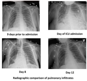
Lungs – at this point it’s beyond much debate, POCUS-enhanced physical examination is vastly superior to radiographs and traditional physical examination. Small effusions are easily seen as well as congestion in the form of B lines. In the case of sub-acute to chronic congestion, as we are not overly concerned with central lesions (not seen with ultrasound), the CXR is of no further benefit.

Peripheral edema – I’ll call this one a tie. Not that much benefit in measuring subcutaneous edema with a probe, except for exact reproducibility, at the cost of time.  😉

The Heart – another no-brainer. Ultrasound wins. With appropriate training, experience, and more important than either, the ability to recognize one’s own limitations.

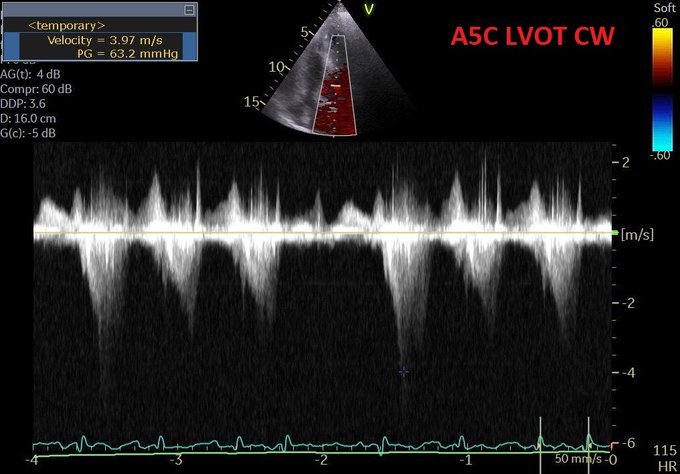
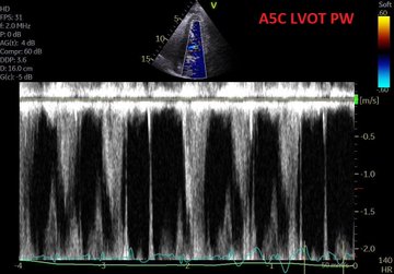
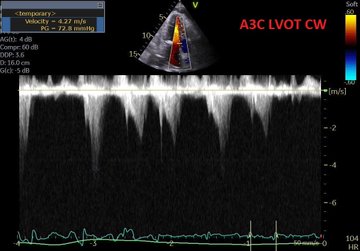
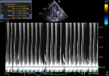
Venous congestion – Now we’re getting to the interesting stuff. So even if for some, it may be the first time hearing about the clinical use of venous congestion markers in CHF, it isn’t new science. In the 90’s, several studies were published correlating portal vein pulsatility, congestion index, as well as hepatic vein doppler pattern with CVP, RV dysfunction, finding close correlation.  In 2016, Iida et al published a great article on renal venous doppler and CHF which I highly recommend reading, and more recently, Andre Denault and William Beaubien-Souligny (@WBeaubien) have been doing tremendous work with portal vein pulsatility and post-op cardiac patients’ organ dysfunction. So the science correlating excessive venous congestion to organ dysfunction is there and is clear.

Why have we not yet widely studied this?

The answer is fairly simple. Prior to the growth of POCUS, there was no single clinician group holding the necessary set of clinical and echographic skills to make this clinical routine. Cardiologists are not all echo-capable, and even those that are would have had little or no experience dopplering abdominal organs and vessels. Radiologists – most of the literature coming from their field – are not pharmaco-clinicians and do not follow patients. Family physicians and internists, likely the bulk of the physicians looking after these patients, largely had not had access to or echo skills. Until now.

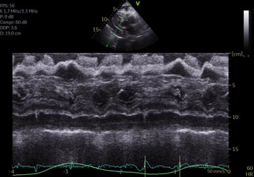
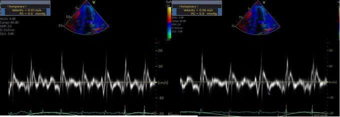
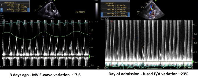
So a quick review:  right-sided failure causes elevated RAP, so everything upstream gets congested. The first echo signal of this is the plethoric IVC (in both axes of course!!!), and an abnormal hepatic vein doppler (which is pretty much like a CVP tracing, just non-invasively) but is that the max? Nope. What is worse is when that pressure transmits thru backwards from hepatic veins to portal vein, transforming a normally monophasic flow with minimal variation into a progressively more pulsatile flow, to the eventual point of being intermittent. And when the IVC pressure transmits across a congested kidney such that the same thing occurs in the renal veins.

Those findings have been well studied and correlate with poor outcomes in CHF.


So what could we do?

What we are doing now is systematically assessing CHF patients in terms of their venous side. What we see so far is that some have full, plethoric IVCs, maybe B lines and effusions, maybe some peripheral edema, but may or may not have those worse markers of abnormal doppler flows, and those who don’t generally don’t have significant organ dysfunction such as renal failure (I discussed this a few years ago in my pre-doppler era in terms of re-thinking common approaches).

So when we find significant portal pulsatility, we diurese aggressively, creatinine notwithstanding. We almost always get an improvement in biochemical markers of renal function within 48-72 hours, with the only really tricky patients being those with severe pulmonary hypertension. More on that in another post.

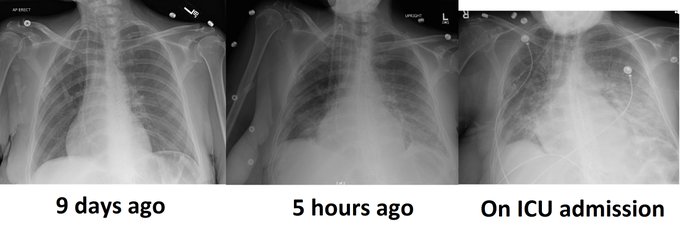
Goonewardena et al had a really great observational study that showed that if CHF patients were discharged with a non-plethoric IVC and significant respiratory variation, they were less likely to be re-admitted. The figure below on the right shows the numbers:

So there is reasonable evidence to suggest a POCUS-guided approach, which we’ll go over in the next post, which should include our revised Advanced CHF Clinic guidelines.

I can already hear the thoughts… “is there any evidence for this?” But those asking that reflexively should first ask themselves “what is the evidence behind the way I assess congestion and manage CHF?”


cheers


Philippe


Refs

Portal vein pulsatility and CHF

Iida et al. article

Beaubien-Souligny and Andre Denault open access article

POCUS, Mythology and Hemodynamic Awesomeness with Jon and Korbin! #FOAMed, #FOAMer, #FOAMus

In Greek mythologyPrometheus (/prəˈmθəs/GreekΠρομηθεύςpronounced [promɛːtʰeús], meaning “forethought”)[1] is a Titanculture hero, and trickster figure who is credited with the creation of man from clay, and who defies the gods by stealing fire and giving it to humanity, an act that enabled progress and civilization. Prometheus is known for his intelligence and as a champion of mankind.[2]

So, fresh from reading Jon’s post, I felt I had to add a bit of nuance in my previous post to what I feared some might extract as a take-home message, even if in fact, we are not that differing in opinion at all – which Jon expressed here:

i agree with ultrasound for finding the uncommon causes of shock. these examples seems to permeate twitter and make ultrasound very appealing. because ultrasound is non-invasive, it makes the risk-to-benefit ratio very low for these uncommon but highly-lethal and treatable causes.

but that needs to be compared to the risk-to-benefit ratio of ultrasound for the more common causes of shock – like ‘non-cardiogenic, septic’ etiologies as seen in SHOC-ED. here, “static’ ultrasound [as per the RUSH and ACES protocols] – per SHOC-ED – appears to be neither helpful nor harmful. your read of the discussion is perfect, but i was depressed because it read as if the authors only realized this ex post facto – study of previous monitoring utensils [e.g. PAC] should have pre-warned the authors …

i will take some mild issue with markers of volume responsiveness and tolerance. you are correct on both fronts – but what the data for the IVC reveals – perhaps paradoxically – is that true fluid responders can have a very wide-range of IVC sizes from small to large and unvarying … this was born out in most of the spontaneously breathing IVC papers [airpetian and more recent corl paper] the sensitivity was rather poor.

the same *could* be true for the opposite side of the coin. a large great vein may not mean a volume intolerant patient. i tried to exemplify how that could be so in the illustrative case in my post. an elderly man, with probable pulmonary hypertension and chronic TR who probably “lives” at high right-sided pressures. nevertheless, he likely has recurrent C. diff and is presenting 1. hypovolemic and 2. fluid responsive despite his high right-sided pressures. portal vein pulsatility *could* be quite high in this patient – but he still needed some volume.

the obvious underlying issue here – which I know you are well attuned to – is that a Bayesian approach is imperative. when you PoCUS your patients, you are inherently taking this into consideration – i know that you are a sophisticated sonographer. my hidden thesis of the post is that if ultrasound findings are followed in a clinical vacuum and followed without really understanding the physiology [which can explain clinico-sonographic dissociation – like the patient in my fictitious case]… disappointment awaits.

Then Korbin Haycock chimes in and adds a level of understanding that I completely agree with but had difficulty in expressing, but which I think is key to understanding the current and future evolution of POCUS. Complex, operator-dependant medical leaps such as laparoscopic surgery suffered with similar growing pains. But I’ll let Korbin shed some light:
I think the issue of POCUS in resuscitation is somewhat analogous to Prometheus’s gift of fire to humanity.
Jon has quite aptly pointed out that if POCUS (particularly a single POCUS supplied data point such as IVC diameter), if used in isolation, without clinical context, and without comprehensive information, is not much better than using a single data point such as CVP to make complex clinical decisions. Multiple factors influence the behavior of the IVC, just as they do with the CVP. Being a dynamic entity, the IVC does have some advantages over a static number like the CVP. However, if considered by itself, the IVC POCUS evaluation will only result in the same pitfalls as using the CVP as a guide to fluid management. If POCUS is applied in such a blunt manner, we are doomed to repeat our previous folly of using the CVP as a guide to fluid resuscitation. I hope I am in the ball park of the core of Jon’s point here, if not as very eloquently stated by him.
Phil is advocating a more nuanced and sophisticated approach to POCUS than what the SHOC-ED trial investigators used to guide management in their study. Most shocked patients presenting to the ED (“Emerge!”) come with a phenotype of distributive shock. Indeed, these were the majority of the patients in the SHOC-ED trial. Any experienced clinician will recognize this syndrome virtually every time, with no more than an “eyeball and Gestalt” assessment from across the room and a set of vital signs. Current dogma is that this syndrome ought to be treated with 30 cc/kg of crystalloids and then to add a vasopressor if the patient’s blood pressure is still low. Given this, there couldn’t have been much difference as to how patients were managed in either group in this study. I however, disagree with this aggressive crystalloid administration approach, as I’m sure many readers of Phil’s blog do as well. What I gather Phil is saying here is, as he insightfully stated in the past, “IVC never lies, it’s just not telling you the whole story.” A complete POCUS gives us (OK, well almost) the whole story. The caveat here is you must know a whole lot about POCUS. Thus the Prometheus analogy. A little information is a child playing with fire.
Someone new to POCUS, with only a novice’s understanding of what an IVC POCUS evaluation means, will probably make the correct assessment of a patient’s fluid status about 60-70% of the time. This probably is only slightly better than an experienced clinician’s non-POCUS judgement. Hardly enough to translate into any meaningful clinical outcome in a trial without a ridiculously large sample size to find a pretty small benefit. But POCUS potentially offers so much more information. LV and RV systolic function, LV and RV diastolic function, SV, CO, SVR, PVR, RAP/CVP, sPAP/mPAP/dPAP, LVEDP/LAP/PAOP, valvular pathology, tamponade, fluid responsiveness (for what ever that’s worth!), RV/LV interactions (both in series and in parallel), EVLW, insight into pulmonary vascular permeability, renal resistive index/renal venous congestion, portal hypertension/congestion, gut flow resistance, and on and on. Most of this information can be more or less determined in less time that it takes to put in a central line in order to get the damned CVP (actually, I do like to know what my CVP is, for what it’s worth). The more data points you are able to collect with increased POCUS skills and experience, the more grasp you have as to what is going on with your patient and the right way to treat them. I would argue that given the information attainable with advanced POCUS skills, POCUS is a no-brainer that will enormously improve not only individual patient outcomes, but effect populations at large, if only the general hospital based practitioner can attain a more than introductory understanding of POCUS.
So, I guess the question is, “how much training is enough training?” I don’t know. Inevitably, POCUS knowledge will incur a bit of the Dunning-Kruger effect as pointed out by Jon’s example of an IVC POCUS fail. But reading Jon’s clinical case example, from the get go, I found myself asking questions that would change may management one way or another with additional information that I could get quickly and easily with additional POCUS interrogation of the patient. Jon pointed this out himself by revealing that the patient has pulmonary hypertension as manifested by the tricuspid regurgitation upon auscultation of the heart. With POCUS, I don’t need to guess what a heart murmur is or how bad it is or even if it is relevant to my patient in this case for that matter. POCUS can tell me it’s TR and it tells me what the sPAP/mPAP/dPAP and PVR is if I care to find out. So if this level of information can be gleaned, for me, no one can argue that POCUS has no merit. But, I’ve spent a lot of time striving to be good at this, just as probably a lot of people reading this have done as well. What about newbies?
Consider: At my main hospital, for a variety of sensible reasons I won’t get into, we decided to train a group of nurses in POCUS in order to evaluate septic patients. They achieve basic training in POCUS and are very competent sonographers with regard to IVC, gross LV and RV function, and pulmonary edema. They are a small group of very intelligent, skillful nurses that are excited to learn all they can. We had them evaluate every septic patient that presented to our hospital, do a POCUS exam, and discuss the findings with a physician. We established some very basic resuscitation endpoints largely based on POCUS findings applied to each individual patient and their POCUS exam. Our severe sepsis/septic shock mortality rates dropped from 35-38% to 8-10% with this program. Our hospital plans to publish this data officially soon for public analysis, but it did make a difference in our experience. That said, my nurses do frequently show me cases where I notice some small detail on their POCUS exam that propmts an additional investigation that alters the plan in management. Also, some of my very competent POCUS savvy residents make errors because they don’t have enough knowledge yet. I’m sure I can make these errors too at times as well, although hopefully less and less so with time.
Here’s my point: Heed Jon’s admonition to look at the big picture and not rely on isolated data points. Be inspired by Phil’s passion for the potential of a good POCUS evaluation. If you only get your toes wet with POCUS, you are playing with forbidden fire. But if you care to look into it further, POCUS opens up worlds to you. By all means, learn all you can about POCUS. Recognize that if you are new to POCUS techniques, there are improtant caveats to each finding, and physiology that needs to be considered with a comprehensive view, some of it may be strictly non-POCUS related information as well. Your patient is unique and only a careful comprehensive consideration of what’s going on with your patient will guide the best approach to your management of their illness. I don’t think SHOC-ED or any other trial for that matter can address the nuances of good individualized patient management. That is up to you.Jon replies:

nice analogy – i think Korbin’s response is appropriate and i look forward to speaking alongside him in May. as i chew on the SHOC-ED a little and try to distill my concerns – i think what it boils down to is this: it’s less about playing with fire – i think – and more about how this fire is brought to the community as a whole. my post on pulmccm was more of a warning to the early adopters [like us] who are planning these trials. imagine 40 years ago:

-the flotation PAC is introduced, a small group of clinical physiologists use it thoughtfully, understand the caveats, the problems of data acquisition, interpretation, implementation, the problems with heart-lung interactions, intra-thoracic pressure, etc.
-these early adopters present their results to the community as a whole
-the physiology of the PAC is simplified
-the numbers from the PAC are introduced into algorithms and protocols and **widely** adopted into clinical practice
-the PAC is studied based on the above and found to make no difference in patient outcome.
-in 2010 a venerable intensivist suggests floating a PAC in a complicated patient and the fellow on rounds chuckles and states that their is ‘no evidence of benefit’

does this sound eerily familiar? is our present rhyming with the past? if the planners of POCUS trials are not careful, i promise you that the same will happen but insert any monitoring tool into the place of PAC. i can very easily visualize a fellow on rounds in the year 2030 scoffing at the idea of PoCUS because trials [SHOC-ED, and future trials x, y and z] showed no difference in patient outcome. is it because PoCUS is unhelpful or is it because the way it was introduced and studied was unhelpful? and the three of us will sound like the defenders of the PAC from 30 years ago: “PoCUS isn’t being used correctly, it’s over-simplified, it works in my hands, etc. etc.”

it’s not PoCUS that’s unhelpful, it’s how we’re implementing it – and i was most depressed when the authors of SHOC-ED appeared to stumble upon this only in the discussion of their paper – like you mentioned phil. imprecise protocols will result in equally imprecise data and the result will be nebulous trial outcomes. we should all be worried.

Korbin adds:

Excellent points Jon. The PAC example is very relevant, as on more than one occasion, I’ve had the argument put to me by some colleagues that essentially how I’m applying POCUS is really no different than the information gleaned from the PAC, and “that’s been shown to not be helpful to outcomes” etc. So, therefore, why do I bother?

Then again, I’ve seen a fair amount of phenylephrine being thrown at hypotensive cardiogenic shock patients after a 2 liter normal saline bolus didn’t do the trick.

You are absolutely spot on when you point out that seeing the big picture, knowing the physiology, and being aware of the pitfalls of isolated data points is important to making the right decisions in patient care.

Furthermore, I agree that when a clinical trial is done that doesn’t consider some of the nuances of all this, and “shows” that POCUS, or any other diagnostic modality for that matter, doesn’t contribute to better patient outcomes, it probably only serves to marginalize a potentially valuable diagnostic tool to an actually astute intelligent clinician.

I’m not meaning by saying this to bash the good intentions of the SHOC-ED trial. To be fair, it’s really hard to design a trial that can take into account all the permutations that are involved in any individual patient presents with, having their own unique clinical situations, hemodynamic profiles, co-morbidities (both known and undiagnosed), etc. POCUS, PAC, transpulmonary thermodilution, ECG, chest x-ray, CT scans, labs, physical exam–these are all merely tools that guide patient care. Albeit some are way more powerful than others. I can image various amounts of uproar if some of these traditional tools were subjected to clinical trials to prove their utility. The argument, if proven “useless” in a study for the oldest and well accepted tools would always be, “put it in the clinical context, and its value speaks for itself.” For me, I’d happily like to make clinical descisions based on information based on an advanced POCUS exam or PAC, rather than interpreting hepatojugular reflux or a supine chest x-ray.

Any diagnostic test requires that the clinician understand the limitations of that test, and understand that the whole clinical scenario must me taken into account. You’ve hit on that, I think, with your argument. This surely has implications when any technology or test is studied.

‘Nuff said.
Philippe
PS These are just the kind of discussions that can change both the way you approach medicine and manage your patients, and these are the ones you find behind the scenes and in the hallways of H&R2018. Don’t miss H&R2019 if you take care of sick patients. It’s the kind of small, chill conference where the faculty will be happy to take a few minutes and discuss cases and answer all your questions (if they can) about acute care.

H&R2019! Final Programme. Register Now! Montreal, May 22-24, 2019! #HR2019

This event is past. It was awesome. If you really wish you’d been there, you can catch most of it here!

And don’t miss H&R2020!

Click here to register!

Registration is open and we have said goodbye to the snail mail process. Fortunately, we are a lot more cutting edge in medicine than in non-medical technology.

We are really excited about this programme, and a lot of it comes from the energy and passion coming from the faculty, who are all really passionate about every topic we have come up with.

The hidden gem in this conference is the 4 x 40 minutes of meet the faculty time that is open to all. Personally I’ve always felt that I learn so much from the 5 minute discussions with these really awesome thinkers and innovators, so wanted to make it a priority that every participant should get to come up to someone and say ‘hey, I had this case, what would you have done?’   Don’t miss it!

CME Accreditation for 14 hours of Category 1.

This programme has benefitted from an unrestricted educational grant from the following sponsors (listed alphabetically):

Cook

Fisher-Paykel Healthcare

GE Healthcare

Maquet-Gettinge

Masimo

Medquest

MD Management

Medtronic

Novartis

Teleflex


The Accreditation is as follows:


Here is the Final Programme:

Final Programme

Wednesday May 22 – PreCongress course

  1. Full day Resuscitative TEE course

FOR DETAILS SEE HERE


    2. Full day Keynotable

    3. Half day Hospitalist POCUS (PM)

    4. Half day Critical Care Procedures (AM)

    5. Half day Brazilian Jiu-Jitsu for MDs (AM)

for more details on these pre-conference courses please see here.


Main Conference Programme: H&R2019 Full Pamphlet

Social Events:

Thursday May 23rd Meet the Faculty cocktail! 1900 – Location TBA – BOOKMARK THIS PAGE!


Register here!

FOR ANY QUESTIONS CONTACT HOSPRESUSCONFERENCE@GMAIL.COM.


Shock Macro and Micro-circulation: Piecing things together. (Part 1) #FOAMed, #FOAMcc


So I have really, really enjoyed the discussions I had with these bright people on shock circulation:

Segun Olusanya (@iceman_ex) Resus Track 2

Rory Spiegel (@EMnerd) Resus Track 3

Korbin Haycock (tell him to get on twitter) Resus Track 4

Jon Emile (@heart-lung)  Resus Track 5


Some take home points so far:

I think that more questions than answers truthfully came out of this, and that is really the best part. But lets see what the common agreed upon thoughts were:

a. the relationship between the MAP and tissue perfusion it quite complex, and definitely not linear. So scrap that idea that more MAP is more perfusion. Could be more, same, or less…

b. you can definitely over-vasoconstrict with vasopressors such that a increasing MAP, at some point, can decrease tissue perfusion. Clinically, we have all seen this.

c. no matter what you are doing theorizing about physiology and resuscitation, THE MOST IMPORTANT IS TO CONTROL THE SOURCE!


Some of the interesting possibilities:

a. Korbin sometimes sees decreasing renal resistive indices with resuscitation, particularly with the addition of vasopressin.

b. the Pmsa – can this be used to assess our stressed volume and affect our fluid/vasopressor balance?

c. trending the end-diastolic velocity as a surrogate for the Pcc and trending the effect of hemodynamic interventions on tissue perfusion.

This stuff is fascinating, as we have essentially no bedside ability to track and measure perfusion at the tissue level. This is definitely a space to watch, and we’ll be digging further into this topic.


Jon-Emile added a really good clinical breakdown:

I think one way to think of it is by an example. Imagine 3 patient’s MAPs are 55 mmHg. You start or increase the norepi dose. You could have three different responses as you interrogate the renal artery with quantitative Doppler:

patient 1: MAP increases to 65 mmHg, and renal artery end-diastolic velocity drops from 30 cm/s to 15 cm/s
patient 2: MAP increases to 65 mmHg and renal artery end-diastolic velocity remains unchanged.
patient 3: MAP increases to 65 mmHg and renal artery EDV rises from 10 cm/s to 25 cm/s

in the first situation, you are probably raising the critical closing pressure [i know i kept saying collapse in the recording] relative to the MAP. the pressure gradient falls and therefore velocity falls at end diastole. one would also expect flow to fall in this case, if you did VTI and calculated area of renal artery. in this situation you are raising arteriolar pressure, but primarily by constriction of downstream vessels and perfusion may be impaired. ***the effects on GFR are complicated and would depend on relative afferent versus efferent constriction***

in the second situation, you have raised MAP, and probably not changed the closing pressure because the velocity at the end of diastole is the same. if you look at figure 2 in the paper linked to above, you can see that increasing *flow* to the arterioles will increase MAP relative to the Pcc [closing pressure]. the increase in flow raises the volume of the arteriole which [as a function of arteriolar compliance] increases the pressure without changing the downstream resistance. increasing flow could be from beta-effects on the heart, or increased venous return from NE effects on the venous side activating the starling mechanism. another mechanism to increase flow and therefore arteriolar pressure relative to the closing pressure is the provision of IV fluids.

in the third situation, MAP rises, and EDV rises which suggests that the closing pressure has also fallen – thus the gradient from MAP to closing pressure rises throughout the cycle. how might this happen? its possible that raising the MAP decreases stimulus for renin release in afferent arteriole, less renin leads to less angiotensin and less efferent constriction. thus, paradoxically, the closing pressure falls with NE! another possibility is opening shunts between afferent and efferent arterioles [per Bellomo]. as above ***the effects on GFR are complicated and would depend on relative afferent versus efferent resistance changes***


This is really, really interesting stuff. So in theory, the MAP-Pcc gradient would be proportional to flow, so if we can estimate the direction of this gradient in response to our interventions, we may be able to decrease iatrogenism. I’ll have to discuss with Jon and Korbin which arterial level we should be ideally interrogating…

More to come, and next up will be Josh Farkas (@Pulmcrit), and I’m sure anyone following this discussion is looking forward to what he has to say. I know I am.

cheers!


Philippe