Pleural effusion: Draining it! (Part 3 of 3) #FOAMed, #FOAMcc

Ok, so once you’ve determined you should drain a pleural effusion, your options are to insert a small catheter (CVC, pigtail, generally 8.5 to 14F) or a traditional chest tube.  CVCs/pigtails should only be inserted when (a) you have ultrasound guidance and (b) the effusion appears to be free flowing and without a great deal of echogenic contents. Why? Because to go in with a sharp needle, as the seldinger technique requires, you need to visualize a safe spot by ultrasound, and because if the effusion is complex with septations and blood/pus, it likely needs a large bore chest tube to successfully drain. For the less experienced, a traditional chest tube insertion is done using blunt dissection and exploration with a finger, such that the risk of pneumothorax is minimal (no needle).

In terms of the pigtail insertion, choose a spot using ultrasound, generally posterior to the midaxillary line to optimize drainage, then freeze, insert the guide needle then guidewire, then dilator and finally the drainage catheter.

Here is a makeshift video showing a pigtail insertion.  I will make another one more focused on the technique, as this one is a bit more focused on the ultrasound. We ca see a large effusion with a bit of atelectatic lung, and also confirm the placement of the guidewire and the pigtail catheter.

Although in this video I showed live ultrasound guidance, in most cases I prefer to pick the spot, mark the skin, and do the technique blind. It is simply faster that way. However in some cases, especially with small fluid collections, I may choose to use live ultrasound guidance.

This is actually a very practical skill to have, as otherwise pigtails are generally inserted by interventional radiology, which implies delays, and more importantly, patient transport and its inherent risks and disadvantages. So for those without experience, I would highly encourage you to seek out an experienced operator and learn this technique.

thanks!

Disclaimer: this description and video is for the sole intent of sharing medical knowledge and does not replace formal training. Do not try this without adequate training and/or supervision.

Pleural Effusion Podcast – physiology and clinical approach! #FOAMed, #FOAMcc

…just for those who prefer to listen rather than stare at a screen…

…and also because I had my sick toddler napping on me for two hours and typing was not an option.

Part 3 with the vid coming in the next couple days, though, apologize for the delay!


Philippe

Pleural effusion in the sick patient (Part 2 of 3): Physiology & Literature! #FOAMed, #FOAMcc

So once your routine cardiopulmonary bedside ultrasound examination has revealed the presence of a significant pleural effusion, should you drain it?

I guess the first real question is actually what is a significant pleural effusion?  As you can imagine, the N=1 principle pops up again, and there is no single threshold answer (e.g.: >750ml  drained and <749ml  ignored), naturally.  So here are the main factors that impact physiologically:

1. underlying global respiratory function – eg how much reserve you have. With healthy lungs, a young adult can usually tolerate complete atelectasis of one lung or even more, which would take 2-3 liters of effusion at least. However, the more compromised gas exchange is, the less atelectasis can be tolerated.  This also encompasses the level of required ventilatory support, including the level of PEEP. Basically, the worse your lungs are, the more significant the effusion becomes.

2. thoracic compliance – which combines chest wall and abdominal pressure, since the diaphragm is thin and easily displaced.  The greater the compliance, the less atelectasis will occur as the diaphragm and chest wall will shift to accommodate some of the fluid. This is very important especially in surgical patients (or anyone with a tense abdomen).

…so there is no number.  Of course, if you take it to the extremes, I think everyone would agree that if you have 200ml,  it’s probably not worth doing, and if you have 2,000ml, it is. But in the grey zone of, say 500-1,000 ml, you have to make a call given the patient in front of you.

Now what does the literature say?

First of all, there is no large study looking at the impact of effusions in generally ill patients (ED/ICU).  There are some small, specific studies showing worse prognosis in some pathologies (eg Legionella pneumonia) but nothing that can really be extrapolated to general ED/ICU patients.

What about in ARDS? Talmor et al, (Surgery 1998, v 123) showed benefit in survival in a small study of ARDS patients.

I think the best overall study was Vignon et al (CCM 2005, v 33) who demonstrated that bedside ultrasound was much more sensitive in diagnosing pleural effusion (CXRs read as normal had as much as 1,000 ml effusions, as well as diagnoses of effusions which were actually consolidations ), but also that it was quite accurate in quantifying (inter-pleural distance >45-50mm at the base correlates with effusions >800 ml).

Usta et al (Interactive CV and thor surg, 2010 v 10) had similar results in spontaneously breathing post op patients with 45 mm (interpleural distance between diaphragm and base of lung) corresponding to about 700ml, although their range was quite wide, and they were using a somewhat impractical sitting position. They actually came up with a formula (16xdistance in mm) to estimate effusion volume.

Sikora et al (ISRN Emergency Medicine 2012) did a good review on the whole thing as well.

Sikora et al review

My empiric observations: I must have put in at least a thousand pigtails or (back in the day) CVCs in pleural effusions over the last 14 years, and my therapeutic goal has always been to drain effusions at least 750 to 1,000 mls. I have drained 350-500 ml in patients in severe respiratory failure a few times, some with apparent benefit, others without. I don’t recall regretting putting one in, but I do regret a few cases of not draining, only to have recurrent respiratory failure post-extubation. Grossly, I would say 750-1,000 or more in a patient with respiratory failure warrants drainage, but 1,000 mls in a CHF patient on nasal prongs can wait for the outcome of diuretic therapy. Of course we are talking about therapeutic and not diagnostic drainage. Under ultrasound guidance with an experienced operator, pneumothoraces should be exceedingly rare.

Bottom Line: start by examining your patient, physically and by bedside ultrasound. Understand his degree of respiratory failure and estimate the size of the effusion and the compliance of his chest.  Then you have to make the call (that’s why we’re MDs) as to whether this particular amount of pleural effusion in this particular patient is worth draining.

Next week:  how to do it video!

cheers!

Philippe

@shanxonline says:

Great post Phillippe and I fully agree that a global assessment is necessary before draining an effusion. With ultrasound we’ve become really good at identifying effusions, but need to understand physiology and especially chest wall/abdominal compliance to understand the physiological implications of drainage vs conservative management.

Pleural effusion in the sick patient (Part 1of 3): Don’t miss it!!! #FOAMed, #FOAMcc

This, in my opinion, is an under-recognized problem when bedside ultrasound is NOT a routine part of examination of critically ill patients. I’m happy to say that as many of my colleagues have been picking up probes, it is somewhat less of an issue now, whereas a couple of years ago I’d often put in 4 or 5 pigtail catheters on day 1 of taking over the ICU.

The first and foremost reason for this is that the portable supine ICU CXR sucks at picking up the small to moderate to, yes, even the large pleural effusion.  Largely owing to the fact that many of our patients have some lung parenchymal abnormalities and to the recumbent position that causes a layering of the effusion, it is often difficult to properly assess the size of a pleural effusion.  Radiologists will usually report the presence of a probable effusion, but quantification is difficult, and physicians not performing routing bedside sonography will often realize the presence of a submassive effusion only on CT scan – after all it isn’t like you can turn and rotate your patient to percuss the shifting dullness, can you?  Not very practical.

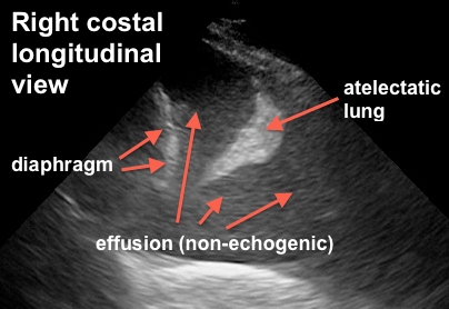
So the following can often be seen:

pleural effusion

this is fairly large, or you might see:

pleural effusion and pneumonia

So the obvious and critical question is: when is it necessary to drain?

There are two elements to this question:

a) for diagnostic purposes: unless the diagnosis is clear (eg CHF, post-resuscitation “michelin man” patient, etc) a new effusion should be tapped.  Panapneumonic effusions, in particular, warrant ruling out empyema unless there is a compelling reason not to.  For diagnostic purposes a 22g needle usually does the trick unless you have frank pus – which generally shows up differently on ultrasound.

b) for therapeutic purposes: effusions are space occupying lesions which compress the lung and result in a variable degree of respiratory compromise, depending on chest wall and diaphragmatic compliance, as well as effusion volume.  The clinical effect is highly variable due to the above as well as the degree of parenchymal lung disease and the degree of PEEP.  In the ICU or ED, a simple way to think about it is that if your patient is in respiratory failure and has a large effusion, chances are that draining it will improve things.  It gets a little more controversial and complicated if you have a patient who is mildly dyspneic with a moderate sized effusion.

Here are a few clinical scenarios I like:

Mr. A is a 65 year old man with CHF, intubated, with large bilateral effusions. He has been aggressively diuresed to the point of his IVC being less than 5mm in diameter.  He has not been able to wean in the last 48 h.

Yes, I definitely drain this fellow. Been there and done that time and time again.  The pleural effusions are essentially the last to resolve (being the most “distal” to the circulation – vs the alveolar tissue itself) and hence can lag and cost a few more days or more of ventilation).

Mr. B is a 47 year old man with pneumonia, breathing spontaneously with a moderate (maybe 500ml) effusion. It appears free flowing and clear, he is afebrile with an improving white count, and mildly dyspneic.

Nah, I skip on this one.  If fever and WBC recur, I do a diagnostic tap to r/o empyema.

Mrs. C is intubated on PEEP 18 FiO2 85% for ARDS due to pancreatitis. She has some degree of intraabdominal hypertension (IAP 18) and has bilateral moderate pleural effusions, maybe 400-500ml.

Yup. She can physiologically benefit from decreased intra-thoracic pressure, both from the ventilatory and the intra-abdominal pressure standpoint (Remember the diaphragm is not a rigid structure so that IAP and ITP are very similar in most cases).

So is there any evidence for this?  Some. And that’s for part 2, coming within the next days. Part three will explain and show my procedure of choice for drainage.

Thanks!

love to hear what other guys’ practices are!   Apparently only about 15% of ICU guys “routinely” drain effusions.

Philippe

COMMENTS

Hi Philippe,
I am very happy to read your post tonight, bacause I am part of that 15% and luckily most of my colleagues are in the same group. I agree in particular when you say that pleural effusions are the last to resolve, being the most “distal” to the circulation. I often find patients, at a certain point during their ICU stay, be not only like a “michelin man” but also (and at the same time!) hypovolemic. I call this situation, when I try to explain it to residents, “empty in full” (maybe in english it doesn’t sound as good as in italian): we are trying with diuretics and some fluid restriction to manage those extravascular fluids that prevent weaning from mechanical ventilation, but often we get the only effect of causing renal failure rather than eliminating pleural effusions. In this case the only way is to drain.
Another important point is that bedside chest x-ray is absolutely useless when you have to discriminate between pleural effusion and parenchimal consolidation, both of them often coexisting in ICU patients.
In our routine we use 14-gauge single lumen CVCs, inserted with Seldinger technique and ultrasound assisted procedure, effective in 95% of the effusions and less invasive than a pleurocath (that we use most of the times for pneumothorax) or a real chest tube, which I keep for blood or traumatic pneumothorax.
Greetings from Italy,
Marco


Glad to hear it Marco!  I started with CVCs as well before we were able to find inexpensive pigtails – email me for info if you want I don’t want to “brand” these things!  They are actually really good because I found CVCs would often occlude. Patient comfort with either is so much more than chest tubes. 

Great point about the “michelin man” who is very “wet” but intravascularly dry, which we see commonly post acute phase of critical illness, especially when physicians are so keen to use crystalloids.

thanks for reading!


Philippe