Test Drive: GE’s New VScan! #FOAMus

I recently had the chance to try out the gen 2 VScan, since my original one, which replaced my stethoscope 4 years ago, suffered from trauma and is currently in a GE operating room undergoing serious surgery.

GE was kind enough to give me a replacement and have me give their next generation a go, so I thought it would be nice to give a little review for potential buyers.

First of all let me put it in perspective. When GE approached me to try the original VScan in 2011, I thought it was a cool toy, but, as an avid sonographer who tended to favour potent devices as opposed to the smaller laptop-based ones, I didn’t expect to fall for the ‘limited’ VScan.  They thought I might.

Turned out we were both right. If I had one of Santa’s elves pushing my ICU’s Aloka Alpha 7 behind me, handing me a clean probe and following me around, plugging and unplugging the awesome but bulky device, I would have no need for a VScan. However, I have yet to hear back from the North Pole, and in its absence, the VScan has been an absolutely awesome and indispensable tool. I’m not joking. My stethoscope has joined my antique medical instruments collection (which includes some really cool ancient metallic tracheostomy tubes btw). I use one of this plastic disposable ones if I really, really want to listen for a wheeze (although I find the degree of expiratory phase prolongation to me a much more sensitive sign of outflow limitation…).

I use the VScan every day, for almost every patient. Bedside ultrasound is an integral part of the physical examination one follow up. It beats guessing.

In my experience, it does the trick about 80% of the time. For more challenging patients, or for a more exhaustive examination, I push that Aloka myself. But when following up patients on the wards especially, having the VScan in my pocket is absolutely essential. Not having it is borderline unethical if I’m dealing with genuinely sick patients.

So the new device has a dual probe with vascular and surface capabilities, which is pretty cool, especially if the physician using it has no other device, as vascular exam is really important for procedures and for lung ultrasound (the gen 1 VScan will see B lines but doesn’t have the resolution to look for lung sliding. Here, I’m using the gen 2 on the wards with a sterile sleeve to drain an intra-abdominal hematoma.

IMG_9049IMG_9052

IMG_9054


So is this a plug for the VScan?  Not really. For bedside ultrasound? Absolutely. Every physician taking care of acutely ill patients should have some form of immediately available ultrasound. Anything else is short-changing your patient of the best care they can get.

My favorite article to illustrate the need to integrate bedside ultrasound in daily examination is the following, where novice medical students with ultrasound blow away seasoned, board-certified cardiologists in cardiac diagnosis:

Screen Shot 2016-03-29 at 10.58.42 AM


Keep in mind that you can still be a great clinician without bedside ultrasound. But you can be a better one with it.


cheers!


Philippe


p.s. thanks to Mr. Pascal Langlois of GE for the gen 2 VScan loaner!


Cerebral & Somatic NIRS (Near InfraRed Spectroscopy) in shock states: tailoring therapy. (PART 1) #FOAMed, #FOAMcc

So I’d mentioned using NIRS to monitor and tailor therapy a few months ago, and promised a more in-depth discussion to come, so here we go.

For this not familiar with the technology or the concept, NIRS measures tissue saturation, predominantly venous. Hence physiologically it is akin to central/mixed venous gases, but localized. Cerebral NIRS found its foothold in the OR with carotid and cardiac surgery, but its use is now expanding. Given typical knowledge translation time of a decade, it should end up joining ETCO2 as a routine vital in monitored units, but probably not soon enough.

So in our unit at Santa Cabrini Hospital in Montreal, we’ve had this technology for about a year (the INVOS system), and have been studying its uses. In this time, three applications have stood out:

  1. Finding the “Sweet Spot” for vasopressors.
  2. Confirmation that therapeutic interventions are hemodynamically appropriate.
  3. Cardiac arrest: CPR adequacy, prognostication and detecting ROSC.


  1. Finding the “Sweet Spot” – I think (hope) that anyone reading this with professional interest understands that pressure does not necessarily equal perfusion.  With that in mind, adjusting vasopressors to a pressure makes little sense, and represents at best a guesstimate of perfusion, which is what we really are after. We can all agree, however, that a certain minimum pressure is required, but whether that is 65, 55 or 45 MAP no one can say for sure.  So the way I like to use it is to establish a baseline and watch the direction of the tissue saturation with vasopressor therapy. If the saturation begins to drop off, we may have reached a point at which excessive vasoconstriction is worsening tissue perfusion, and that inflexion point may represent the upper beneficial limit of the vasopressor – this may happen to be under 60 or 65 of MAP.  However, it is key to understand that this inflexion point is reflective of the current state of hemodynamics, such that a change in volume status or cardiac output, in one direction or the other, would likely change the position of this physiological point.  For example,  a volume depleted patient may reach a decreasing tissue saturation point at 55 MAP, but, once volume replete, may reach a higher MAP of 65 or above before a drop in saturation is seen.  Conversely, a patient whose best tissue saturations were around 65 MAP who suffers an MI and sudden drop in cardiac output may now see his perfusion compromised at that same MAP, which would now be achieved with a greater vasoconstriction, less cardiac output and consequently, poorer flow… I posted a case discussion which illustrates this.
  2. Confirmation that therapeutic interventions are hemodynamically appropriate – I feel this is really important. When a patient’s life is literally on the line, and knowing that our interventions are seldom without potential nefarious side effects, it is poor medicine to be introducing a therapy without having some form of monitoring – preferably multiple – that we are headed in the right direction, or at least not making things worse. Of course, we already do this – with BP, sat, lactate, CCO, ultrasound, ETCO2 – but I think using a realtime measure of tissue saturation adds to this. It is also my firm opinion that integrated, multimodality monitoring is necessary – at least until someone develops some form of mitochondrial monitoring which tells us that the cytoenergetics are sufficient to survive. Until then we are stuck with surrogate markers and many of them (e.g. lactate) are the result of complex processes that preclude them being a simple indicator of perfusion adequacy. For instance, when giving a fluid bolus/infusion – after having determined that the patient is likely fluid responsive AND tolerant – one should expect to see an increase in ETCO2 (other parameters being constant), an increase in CO, an increase in NIRS values. The absence of such response should make one reconsider the intervention, because without benefit, we are left only with side effects.

Here is a patient’s cerebral (top) and and somatic (thigh – bottom) and CO values. This patient had an RV infarct and was in shock.

IMG_7948IMG_7946


Following initiation of dobutamine, this is what occurred:

IMG_7951IMG_7949

Given that we cannot always predict the response to an inotrope – depending on the amount of recruitable myocardium, it is reassuring to see an improving trend. This enabled us to decrease the vasopressor dose significantly.

Note that, so far, and unless some good evidence comes out, I don’t use a goal value, and so far, I have not identified a value that is predictive of prognosis. However, downward trends usually bode very poorly. For instance, I had a severe chronic cardiomyopathy patient whose cerebral saturation was 15%!!!  But more surprisingly, she was awake, alert and hemodynamically stable. Adaptation.

Part 2 and the stuff on cardiac arrest coming soon!

Please, anyone using NIRS in shock, share your experience!


cheers


Philippe

Volume responsiveness and volume tolerance: a conceptual diagram. #FOAMed, #FOAMcc, #FOAMus

So I know I’ve belaboured the point about the difference between volume responsiveness (i.e. will there be significant increase in cardiac output with volume infusion) and volume tolerance (is the volume I am considering giving going to have nefarious consequences), because in my opinion, the focus has been – rightly so to some degree – to look for an accurate way of discerning responsive patients from non. Of course this is absolutely necessary, as one does not want to give volume if it will not have any benefit, but the too-common corollary to that is to automatically give volume to those who are responsive.  Here is an earlier post about this:

Fluid Responsiveness: Getting the right answer to the wrong question. #FOAMed, #FOAMcc, #FOAMus

So in discussing with a bright young colleague yesterday, Dr. St-Arnaud (@phil_star_sail), I realized that there may be a common conception that physiologically, the relationship between the two may be the following:

Screen Shot 2016-02-21 at 9.03.01 AM

This would mean that it is safe to give volume until a patient is no longer volume responsive, and even perhaps a bit more. Alternately, the two may be closer:

Screen Shot 2016-02-21 at 9.02.27 AM

This would mean that once can go just till the point where the patient is no longer volume responsive.

Either one of these scenarios would be awesome. That would mean that by using any of the flow or volume variation techniques, arterial or venous, we could pretty much remain safe.

However…

While the above may hold true for healthy subjects, I would contend that in sick people (which is who I tend to deal with, especially when resuscitating shock), that the more likely physiological relationship is the following:

Screen Shot 2016-02-21 at 9.03.28 AM

Hmmm… That would mean that assessing for volume responsiveness would only tell you that there would be an increase in cardiac output, but absolutely nothing about whether it would be safe to do so.

This concept is not a new one by any stretch of the imagination. It’s inferred in the diagnosis of “non-cardiogenic pulmonary oedema.” So what causes this shift? Here:

Screen Shot 2016-02-21 at 9.25.50 AM

So, how do we figure out where the point is? Sorry to say there is no answer that I know of. My friend Daniel Lichtenstein uses the FALLS Protocol (identifying the appearance of B lines during resuscitation) which is the least we should do, but I suspect that at that point, we have already overshot the mark. My adopted mentor Dr. Andre Denault (@Ad12andre, in addition to IVC, has identified portal vein characteristics including pulsatility (lots of stuff in press) to show that the viscera are at risk, but as of yet there is no simple answer. CVP value? Please. CVP tracing morphology? Maybe.

No simple answer. No one-size-fits-all velue to look for. Clinical integration.

In my opinion, one should not, in sick patients, seek to volume resuscitate until the point of no-volume-responsiveness. The old adage of “you have to swell to get well” likely kills a few additional patients along the way, just as much as under-resuscitation. I plead guilty for over-resuscitating patients for years before realizing that being on the flat part of Frank-Starling is 100% a pathological state.

Love to hear your ideas and comments!


Jon-Emile Kenny says:

I like your graphics, it makes the concepts tangible. I think we should try to integrate ‘volume status’ into this framework as well. A physiological purist might say that as soon as you are ‘hypervolemic’, you are volume intolerant, because hypervolemia is an abnormal state which should always be avoided. A functionalist might say that you become volume intolerant as soon as you have physiological embarrassment of any organ system – but how is this determined? My gut is that by the time there are B-lines in the lung, you’ve gone too far. By the time there is abnormality of splanchnic venous return, you’ve already gone too far. I am more of a purist, so in my perfect ICU, I would perform q4-6 hour radio-labeled albumin studies to determine the patient’s true plasma volume. In health, the normal blood volume is about 80 mL/kg [thus, once you’ve given a 70kg man 5 L of NS, you’ve almost certainly replenished his vascular volume]. The moment that the blood volume becomes > 95% the norm, I would call the patient volume intolerant and stop volume expansion and focus on venous tone with pressors, cardiac function with inotropes, etc. To me, this makes the most sense in the pure Guytonian world; if you keep flogging a patient with litre after litre of fluid and the patient’s BP remains low, you are missing something – volume is not the answer – regardless of what an ultrasound shows you:
1. trouble shoot the venous return curve [i.e. too little blood volume, too little venous tone, too high resistance to venous return]
2. trouble shoot the cardiac function [i.e. poor rate, rhythm, contractility, valve function, biventricular afterload]
If you need some objective measure of blood volume before you can call volume status optimized before moving onto the next problem to fix – that’s a radio-labeled albumin.
Maybe I’m crazy.cheers

Jon

Thanks for commenting Jon!

I totally agree, if we knew each patient’s normal blood volume, that would be a starting point.  And of course, that would prevent the over resuscitation of a very dilated and compliant venous system (small IVC on ultrasound). Let us know if you figure out a practical way to do that!

It’s too bad that extravascular lung water doesn’t seem to have panned out – not sure why exactly.


Philippe

Bedside Ultrasound & Intracavitary Thrombolysis: Using the Ajmo sign. #FOAMed, #FOAMcc, #FOAMus

So as a follow up to a recent post about intra-abdominal thrombolysis, here is a little pearl from a colleague and fellow ICU physician, Dr. Ian Ajmo.

The Ajmo sign refers to the visualization of injected contrast in a fluid cavity, used to confirm proper position of a drainage catheter in an effusion or ascites.

In the case of a separated effusion, it is often difficult to determine if the septations are truly divisive of the fluid, or just a network of membranes that remain communication and that a single catheter can drain. I have seen both cases with similar echo graphic appearance.

However, the Ajmo sign can be used to determine of the catheter is likely to drain the bulk of the collections, or if the use of thrombolytics should be considered:


In this case, we can see that the agitated saline fills only one of the cavities with little or no spillover into adjacent pockets of pleural fluid. This is a case where thrombolytics can be considered. Hence, this consideration can be done early using bedside ultrasound, rather than follow up CT scans (notoriously poor at seeing fine separations).


cheers!

Philippe

Bedside Ultrasound for neuro-assessment in the ICU: another interesting case! #FOAMed, #FOAMcc, #FOAMus

So a woman in her 40’s was discovered unresponsive at home and emergently brought to hospital. She was known for severe rheumatoid arthritis and had had two CVAs in the past. She had unfortunately suffered from distrust and noncompliance.

An emergency cricothryrotomy was done for a difficult airway which, due to difficulty ventilating (no cuff in our cric kit) was exchanged to a 6.0 ETT over a tube exchanger. Unfortunately our CT scan was up for maintenance, so the CT head was delayed.

Our working diagnosis was another CVA with an aspiration pneumonia.

So of course we had to pick up the probe and take a look:


778_20160121_OEIL_0008


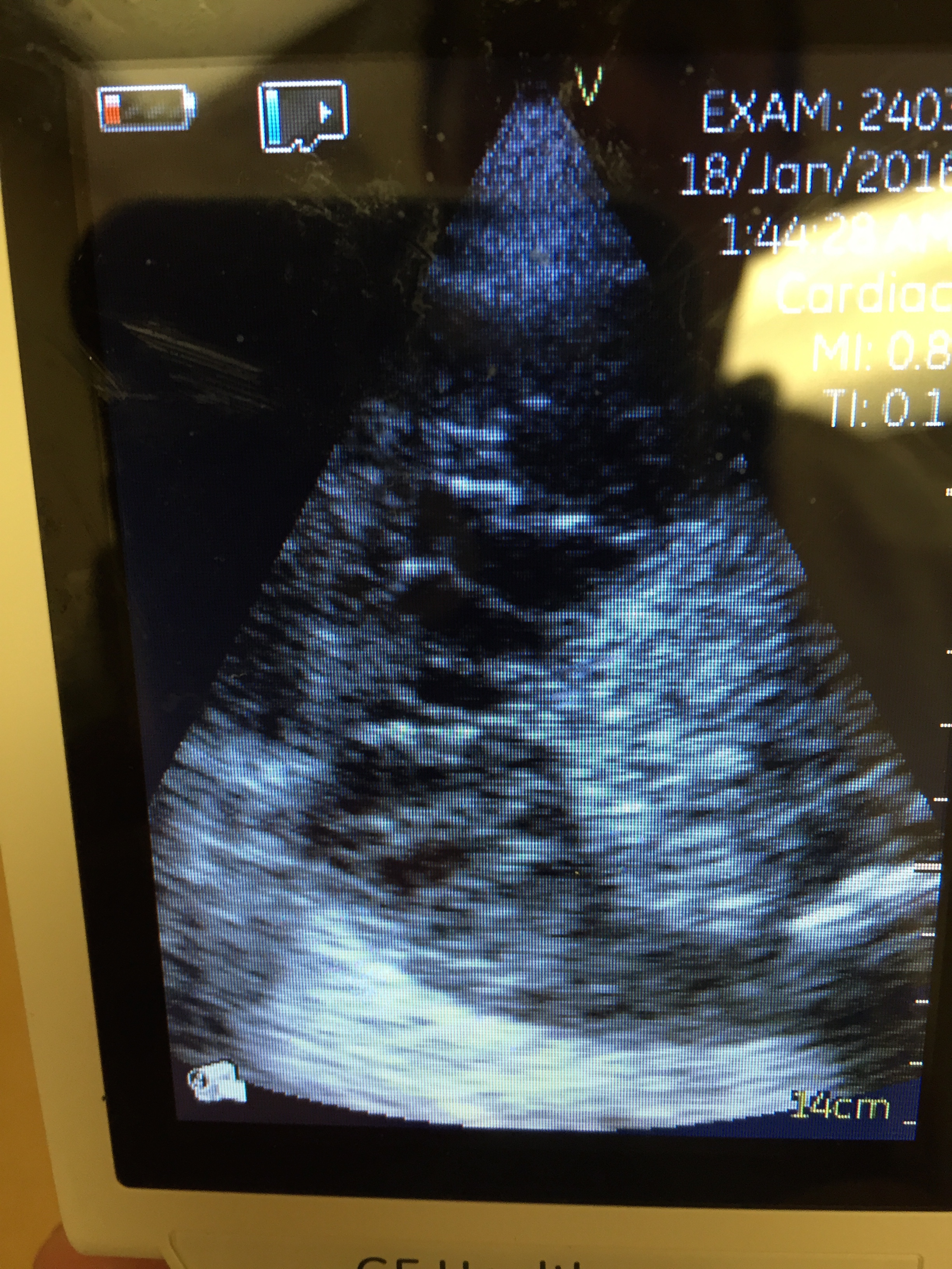
So the optic nerves are dilated significantly (bilaterally), suggesting an ICP over 20 mmhm at the very least.

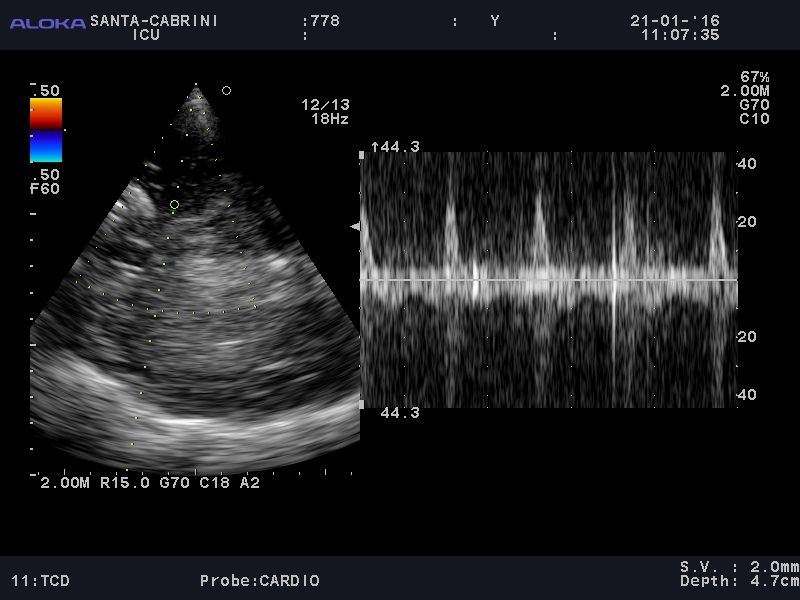
Next the TCD waveform (from the left temporal area):

778_20160121_TCD_0010

It was fairly difficult to find a good vessel, and likely at a bifurcation hence a narrow and bidirectional spike on doppler. The signal noise made it difficult to look clearly at diastolic flow and make a definitive call of circulatory arrest.

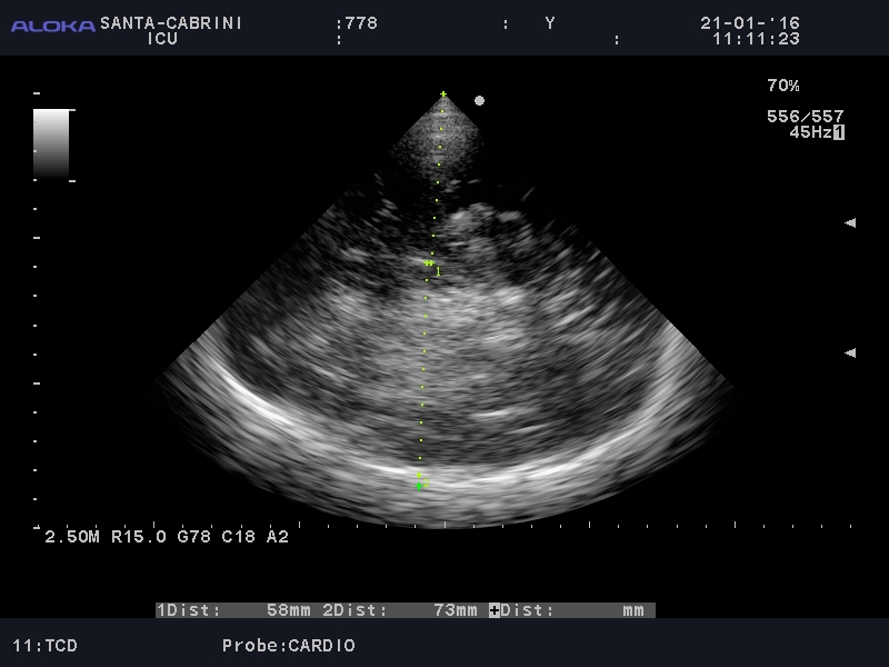
However, there is a clear asymmetry in the hemispheres (not due to gain difference), with the distal hemisphere (RT) showing increased echogenicity:

778_20160121_TCD_0005

Also, there is a midline shift:

778_20160121_TCD_0007

This suggested an acute and severe process in the right hemisphere. A few hours later, the CT scan showed the following:


After family discussion, we withdrew therapy.

So one of the questions a colleague asked is whether I would have been comfortable making the call of an acute and severe process at the bedside and deciding to withdraw therapy, and at this point, I would have said no, in this particular case where the TCD signal wasn’t quite clean enough, and in a center with CT scan availability. I could, however, see a clinical scenario where, with a better TCD signal, I might do so, especially if I were in a remote area.

cheers!


Philippe


So to answer my colleague’s question about “calling it,” I reached out to the one who first started me in CNS ultrasound, Dr. Robert Chen (@ottawaheartrob), who had this very, very insightful comment (originally on his blog)

 
Can you “call it” with CNS POCUS?
1/30/2016
A dear friend and colleague wrestled with a difficult situation.

https://thinkingcriticalcare.com/2016/01/24/bedside-ultrasound-for-neuro-assessment-in-the-icu-another-interesting-case-foamed-foamcc-foamus/

A patient, who despite chronic diseases had distanced themselves from the medical system. Found collapsed, a long and hard fought resuscitation ensued. Airway challenges, ventilation and oxygenation challenges had to be overcome as part of a prolonged code. After Return of Spontaneous Circulation (ROSC), attention turned to the neurologic exam. Beyond the clinical history of prolonged cardiac arrest with the complicating hypoxaemia, the clinical exam was non-reassuring. Technical factors delayed access to CT scanning of the brain.

Optic nerve sheath ultrasound (an anatomic assessment) suggested raised ICP. Transcranial Doppler suggested the physiology of raised ICP. 2D grey scale ultrasound imaging of brain demonstrated asymmetry (yes, the team was THAT good). The question: should one stop resuscitation based on CNS POCUS alone?.

“Calling it” in resuscitation has a long history of being based on the clinical history and clinical examination alone. Think of the many “code blues” that you have attended. Many efforts have terminated with the team having decided that the patient could not be resuscitated without any imaging modalities employed, point of care (POC) or other. “Calling it” with POC ultrasound (POCUS) starts its history in the emergency department which eventually became the American College of Surgery’s Committee on Trauma (ACS COT) codified Focused Abdominal Sonography in Trauma (FAST). Patients were sent for laparotomy or subxipoid pericardial window based on POCUS alone. POCUS, in great urgency offered the clinician improved access to diagnosis and thus improved access to resuscitation direction for the patient.

What about “Calling it” for brain injury? Does the faster access to resuscitation mean something in this situation?

CT scans of the brain are the standard of care in this situation. POCUS has never meant to replace standard imaging modalities (echocardiography by cardiology, abdominal imaging by radiology, vascular Doppler, etc.) but rather to augment the physical examination, screen for time critical injuries and offer easy repeatability particularly in time sensitive situations. Which POCUS element is important in this case? None and all.

Would one stop CNS resuscitation without knowing the mechanism or resuscitatability of the CNS injury without a CT scan? Even in the hands of a vascular Doppler lab, transcranial Dopplers are supportive tests at best for neurologic determination of death (as opposed to ancilliary tests: 4 vessel angio, CT angio or scintigraphy). Stopping resuscitation at this phase (ROSC) based on POCUS would put the patient into a palliative mode as opposed to stopping a code wherein the previous diagnosis of death and non-resuscitatability is acknowledged.

Still, should they have “called it”? POCUS does another wonderful thing. It asks the clinician to touch their patient. Rather than being the acute care quarterback (often recording the nurses vital signs rather than performing their own) who orders “STAT ECG, CKs, troponins, and get cardiology here to do an echo” the POCUS able clinician spends a few more seconds examining and speaking to the patient, often offering the patient more clinical information quickly.

POCUS in this case in the hands of a caring clinician may do something different: when speaking to family they would be able to express that patient had perhaps already declared their intention through distancing themselves from the medical system. As well, they would be able to say “After I examined her, I was so worried that I did my own testing. I did a focused brain ultrasound which makes me very concerned that something terrible has happened: a stroke, a bleed, a tumour, something bad. What I would have normally done is order a CT scan of her head and then possibly call a neurosurgeon. What would she want me to do?”

POCUS brings us closer to out patients. We examine them for longer. Describing POCUS results and their significance in a contemporary fashion to the patient and/or their loved ones forces us to communicate more.

Stop CNS resuscitation without tomographic imaging in Canada? No. Understand CNS anatomy and physiology better through POCUS? Sure. Become a better CNS resuscitator as a result? Probably. Communicate better to patients and families at the end of life through POCUS? A strong possibility.

Intra-abdominal thrombolysis for septated SBP: a case. #FOAMed, #FOAMcc

So we had an interesting case this week of an alcoholic cirrhotic fellow in his 60’s who was admitted with SBP, septic and in respiratory failure. A pigtail had been inserted (RLQ) a few days ago successfully draining 3 litres of cloudy ascites which grew morganella and e.coli. Obviously he was treated with antibiotics.

When I took him over on a monday morning he was hemodynamically stable but with a distended and tense abdomen. A quick look with bedside ultrasound revealed significant but highly septated ascites, and the pigtail had been draining little in the last 24 hours.

IMG_7538

We decided to insert a second pigtail in this area, which immediately drained only 30-40 cc, and decided to use TPA to loosen things up a bit (we used 2mg TPA in each pigtail).  In the following 12 hours, the original pigtail drained 700 cloudy cc’s and the new one, 1,000 cc’s. We repeated the TPA for two more doses but little more came out. No bleeding, and he was on prophylactic anticoagulation.

Just thought I’d put the case out there to add to the intra-abdominal thrombolysis data, which is substantially less than the pleural, and, as usual, to show how routine use of bedside ultrasound reveals things you’d either never otherwise see (loculations on CT???  Naaaah.), be guessing about, or have to wait and move your patient to CT.


Cheers!


Philippe


Here is a nice little review on lysis: sir04264

Bedside Ultrasound-Assisted Procedure: Hepatic abcess drainage. #FOAMed, #FOAMcc

Hi,

Apologies for a long hiatus. Thought I’d share a case from last night. A 54 year old man had been admitted with e.coli sepsis complicated by portal vein thrombosis and multiple hepatic lesions a few weeks ago. A follow up scan by the hospitalist showed the following:

Yup, nasty. So our ICU Outreach service was called (we do all manners of procedures on the wards/er) and it happened to be me.

So 10pm I make my way with all the necessary gear (not much you can’t get done with ultrasound and caffeine!):

IMG_4242

Here is the clip:

So this is a synthesis of several US loops. The first ones simply show the lesion, which under US is clearly fluid – movement well seen with respiration/pulsation. Next you see the associated ascites and a quick peek at a subxiphoid view of the heart.

You then see the procedure itself, with a needle insertion (purposely jerky for visualization’s sake), and, following a 3 way stopcock connection, gradual drainage of the abcess.

IMG_4246

I chose to hand-drain it in this case to avoid possible blockage of the tube if simply left, since it was a small 8.5 french pigtail catheter (better for comfort). You can see that the access cavity was essentially obliterated. 400 ml or so drained:

IMG_4247

So technically this was very simple, however the one important teaching point is to pick an inferio-lateral approach, as an easier but more treacherous one – simple lateral – might result in going thru the pleural space because of the lateral costodiaphragmatic recess which extends quite inferiorly. So when picking the entry point, it is important to make sure it is below the diaphragmatic insertion. Otherwise the potential to seed the pleural space with abcess content is there. This would be sub-optimal.

The advantage of bedside ultrasound? Quick and easy drainage during the weekend when interventional radiology isn’t readily available.

cheers!

Philippe

Enteral Fluid Resuscitation (EFR): Third-world medicine in the modern ED/ICU? (ORT part 2) – #FOAMed, #FOAMcc, #FOAMer

Screen Shot 2015-02-10 at 7.15.16 PM Enteral Fluid Resuscitation in the ER/ICU? For those who did’t come across it, part 1 of this series can be found here: http://wp.me/p1avUV-e8 So back to bringing the basics back to our ultra-tech world… Can I actually use this field technique in my bright and shiny ICU? Can I use oral hydration as a cutting edge therapy in my life-and-death patients? Sounds strange. Sounds like I should be using a precise device which lets me know exactly how much fluid has gone into my vascular space, because that’s where I want it to go, and I want to control exactly the composition of my serum electrolytes, etc, etc.  We like to control. But do we really? We actually have absolutely no idea how much of a fluid bolus remains intravascularly, in any one patient.  It will depend on his/her pre-existing venous filling, his serum protein levels, the integrity of the glycocalyx, and probably a few more things we don’t even know yet.  And as I rapidly distend atria, I release ANP which damages my glycocalyx further. Hmmm… As I mentioned in the last post, the only way fluid enters our vascular space is via the endothelial cells at the level of the GI tract for the most part. All “venous access” is iatrogenic. I do believe that the endothelial cells, by and large, will do a better job – in concert with the kidneys and rest of the blood cells – of controlling the plasma than we will, if given the chance. What logically follows is that, in the presence of a functional gut, I can consider using Enteral Fluid Resuscitation – that is, giving fluid for hemodynamic purposes, not just “maintenance,” by an enteral tube of some sort. So what could I give?   What’s in it? The current reduced osmolarity WHO/UNICEF formula contains approximately the following: Screen Shot 2015-02-10 at 7.24.28 PM So, lets take a closer look at the players: 75 mmol/l of sodium, 75 mmol/l glucose, some potassium and the rest basically to balance the electroneutrality. The whole thing hinges on the glucose-sodium cotransporter, which drags sodium and water in along with the “desired” glucose.  Optimal water absorption takes place with Na between 40-90 mmol/l, glucose 110-140 mmol/l, and an osmolality around 290.  A higher Na may cause some hypernatremia, and a higher osmolality may result in water loss. Here is our friend the enterocyte illustrating just how this kind of solution will allow sodium absorption: Screen Shot 2015-02-10 at 7.31.54 PM   Do-it-Yourself Enteral Fluid Resuscitation Solution: So I’ve got a neat DIY option if I don’t want to break out the powder and start mixing in the middle of my unit: 0.45% or 1/2 NS plus 30 ml of of D50 would give us Na 77, Cl 77 and glucose 74, with an osmolality of 228. Pretty close. That’s what I’ve been using. How much? Well, I like the slow and gradual. Some of the rehydration data out there supports some pretty huge amount of fluids, but this has been done mostly in healthy but dehydrated athletes – not the case for most of our patients. I’ve been going with 250ml every 1-2h, as – for now – an adjunct to IV fluid therapy. This is conservative and completely arbitrary, but essentially a glass every hour or two certainly doesn’t seem excessively taxing. Who can I give this to? You do need a functional gut, so for now, my criteria are (1) essentially normal abdominal exam, (2) obviously no recent bowel surgery, (3) a patent and functional gut as far as I know, (4) no ultrasound evidence of ileus or gastric distension. But how can I be sure it’s going in the right place?  I can’t. Just like I can’t be sure my IV fluids are staying in the right place. But I do check – IVC ultrasound (gross but better than skin turgor!), urine output, HR, BP, etc. None of those are perfect as they are all multifactorial, but that is the nature of the game. The other thing I check is gastric distension by bedside ultrasound every couple of hours – obviously, if I’m just getting a fluid filled stomach, there’s no point, and eventually harm may ensue. When should I stop? Whenever you clinically decide you don’t need/want any further fluid resuscitation. As far as I am concerned, might as well stop the IV infusions first and have the enteral going after – in the end, you are hoping your patient will go back to drinking and not require a PICC line for discharge, aren’t you?  So you stop when the patient does it on his or her own. I’d really like to know if anyone out there is doing something like this. It would be great to compare notes and evolution. Drop a line!   cheers, Philippe

Lung Ultrasound Day for Internists 2015: ASMIQ/CCUS, Montreal, May 29th 2015.

In collaboration with the Quebec Association of Internal Medicine Specialists (ASMIQ), and featuring none other than Dr. Daniel Lichtenstein (the original pioneer of bedside ultrasound and the first to truly do and develop lung ultrasound), we’ve put together a really good day of lectures and workshops. As a bonus, my good friend Dr. Haney Mallemat (@criticalcarenow) will be giving us a hand with workshops and maybe even a special lecture! Take a look:

ASMIQ 2015 Lung US Program

This takes place in Montreal, at Santa Cabrini Hospital, and places are limited, so anyone interested should register promptly at http://www.asmiq.org.

Do note that most lectures will be in french!

thanks,


Philippe

Bedside Ultrasound Case: Look Left and Right! #FOAMed, #FOAMus

So I get a patient in the ED who had chest pain and a decreased LOC, vomited and got intubated. I see this elderly (88 yrs old) gentleman a couple of hours after presentation, after basic management including some plavix and heparin for a mildly elevated troponin.

Of course, by now you all realize that a rapid CUSE (Critical UltraSound Examination) is what I start with, after an ultrabasic history.

So my first couple of views show a more-or-less normal IVC, and here is the parasternal long axis:

Anything exciting here? Not really, nothing to hang your hat on at a glance.

Ok, so thanks to FOAM, I recently decided to add the right parasternal view to my regular exam, both to look for lung sliding (I admit I sometimes skip this when not specifically looking for pneumothorax) but also to possibly see some right sided pericardial abnormalities, etc… Here is what I see:

Hmm… A large, vascular structure that seems to have two lumens… a flap? Back to the patient exam, and the left toe is upgoing  and seems more flaccid in the left upper extremity…

Lets creep up the vascular path to the neck vessels:

Here, we can clearly see that most of the carotid lumen (lower right) doesn’t have any flow. That’s suboptimal. In fact, only a small crescent of flow between 3 and 6 o’clock is seen.

Here is the CT:


So here we can clearly see the dissected ascending aortic aneurysm that extends into the right carotid artery.

Due to advanced age and dismal overall prognosis, support was discontinued after discussion with the family.

I thought this would be a great case to share due to the fact that it could have been an initial bedside diagnosis, but I have to say I consider it fortuitous that I happened to look right, then up – which I easily could not have done. Not that it made any difference in this case, but on the next one, it just might!


Thanks FOAM!


cheers


Philippe