Enteral Fluid Resuscitation? The WHO to the rescue in the ED/ICU? (ORT part 1) #FOAMed, #FOAMcc, #FOAMer

So something has been trotting around my head for a few months, and it actually stems from a small and not-so-proud moment I experienced during a conversation with my wife, while she was still a resident.

She was telling me some of the stories of the day, and how one of her supervisors who had a mixed outpatient and ED practice, always pushed them to use PO fluids, get rid of IVs and get the patients home.  I kind of scoffed, in a sadly typical acute care physician mode, saying how you had to be a bit more aggressive and give them IV fluids to revert their dehydration a bit faster.

Then I caught myself. Hmmm. What exactly am I saying this (con brio) on the basis of. Knowledge, or belief?    I tried to find knowledge but came up woefully short. It seems I’m doing this out of habit, what I’ve seen/learned/believed in the two decades since someone handed me an MD degree. Damn.

So, I do believe in evolution. We have evolved platelets to stop bleeding, fibroblasts and osteoblasts that can fix bones, white cells that go mop up the messes, and all kinds of other good stuff.  One thing we do NOT have is small openings in vascular structures that allow unprocessed, man-made fluids directly into the bloodstream. We make these. We insert tubing into normally sterile environment and infuse a vast number of medications directly into this fragile matrix of cells and organic colloid – with the best of intentions.

In our physiology, however, the ONLY way fluid ever enters the vascular spaces is by diffusion from the outside of the endothelial cell into the lumen, molecule by molecule and ion by ion.

So let me seemingly diverge for a bit…

Screen Shot 2015-02-09 at 12.05.58 PM

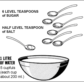
Prior to the 1970’s, restricting oral intake was a “cornerstone” therapy of diarrheal illness, due to the pervasive belief that the GI tract needed time to heal and recover before resuming normal function. This was felt to be crucial. Hence, only IV therapy was used (in developed countries), and in the underdeveloped world, the death toll was appalling – especially among children.   In the 40’s, Dr. Darrow of Yale started actually studying the GI tract fluid and electrolyte issue, and advocating oral rehydration with mixed fluids. He was able to bring infant mortality radically down in his practice, but it would take over twenty years before a groups started to formally look at this in the 60’s.  Finally, in the late 70’s, the WHO pushed this out into the field, and the childhood worldwide mortality from acute diarrheal illness dropped by over 70%, from over 5 million deaths a year to a bit over 1 million – at that time.

Oral Rehydration Therapy (ORT) is now felt to be one of the most significant advances in modern medicine. Compared to that impact, all the critical care and cardiology trials are about as significant as a drop in a bucket. We’re not talking about composite end points and subgroup odds ratios of 0.85…

For a great review on this check out The History of Oral Rehydration Therapy by Joshua Nalibow Ruxin (google it).  A great story of science and humanity, good and bad.

So, back to 2015 ED/ICU’s.

Screen Shot 2015-02-09 at 12.06.26 PM

The question now becomes the following: why – in the presence of a functional gut – do I choose to entirely rely on non-physiological IV fluid resuscitation?

I can already hear the roars and the outrage and the cries of heresy.  And heresy is certainly what this is (Heresy is any provocative belief or theory that is strongly at variance with established beliefs or customs – Wikipedia). But that doesn’t make it wrong.

So I would ask everyone – particularly the naysayers, to examine their knowledge and see if they actually have any at all that supports the strong conviction that IV fluids are the way to go in ALL cases (my N=1  principle precludes going for the one-size-fits-all therapeutic approach).

Now everyone agrees that, once patients are better, they should be on feeds with little maintenance fluids. I don’t think many will debate that. So that should be the basis to wonder whether, in the presence of a functional gut, a variable proportion of fluid resuscitation in acute illness should be enteral…

I’ll let everyone digest that.

Comments more than welcome.

More to come in Part 2.


cheers!

Philippe

Post-Arrest BP Study by Young et al (Resuscitation) – interesting & important, but not yet an N=1 answer! #FOAMed, #FOAMcc

Happy New Year to all!

So trying to catch up on some reading, here is an interesting paper I came across. Young et al did a retrospective study on post-arrest BP, in an attempt to answer the very pertinent and important question as to whether or not a higher MAP may confer better neurological recovery, which is a very sensical hypothesis. After all, a brain with potential swelling, both of tissue and endothelium, may “need” a higher BP. Some societies have advocated for a higher MAP than is usually targeted (i.e. 65) and in studies this has been anywhere from 60 to 100. In their particular institution (Vanderbilt) the protocol aimed for 80-90.

Here it is:

Young et al RESUS

So what did they find?

Basically, they were unable to demonstrate that a higher MAP – in this case defined as achieving 80 mmhm – improved anything, with a follow up to 3 months. There was also no increased mortality related to the use of vasopressors.

So, why might this be? Well, I think there are a couple of important principles to review, especially for the novices reading this.

1. Pressure does not equal flow. The relationship between pressure and flow is a complex one and depends on the interaction between the pump (CO) and the circuit resistance (SVR). Pressure rises when resistance is increased, output is increased, or both. If resistance is increased without increasing output – or by a disproportionate increase in resistance vs output, flow decreases. The effect of vasopressors such as norepinephrine is complex, with both vasoconstriction and increased cardiac output (both via beta stimulation and via increased venous return), and depends on volume status, alpha sensitivity and the recruitable cardiac reserve.

So…? This means that on the surface, a BP number tells you little about flow. The same MAP may represent a highly vasoconstrictor, low-flow state, or a normal flow state. Obviously, a certain minimum pressure is required, to drive the flow from artery down thru the capillaries, but what that number is is unclear. So when looking at any study using simply MAP without another assessment of flow, one cannot draw a conclusion that improving hemodynamics may not help the situation.  How does one assess this – in all likelihood  an integrated approach using ultrasound (volume status, cardiac function), tissue saturation (cerebral/somatic oximetry) and possibly other technologies, including simple physical exam looking at skin mottling.  This type of information could categorize patients into flow categories and make results much more interesting and applicable.

Note that this isn’t really criticism on the authors – it would be impossible to do this on a retrospective study, but simply food for thought for further studies to come.

2. The N=1 principle: remember that we are never treating hundreds of patients at once, and we do not have to decide what is best for most (which is what an RCT generally answers) but what is best for the one patient we are treating.  Hence, looking at any one patient and saying that the target BP should be 65 vs 80 based on this study is incorrect.  What we should be saying is that aiming for a higher MAP may not be necessary if we feel that the patient is well perfused at 65. How each of us figures that out will depend on individual skills and available technologies, but to simply aim for 65 without further thought and assessment is relinquishing your MD in favour of the printed word, essentially what any paper protocol could do.

In the next post I’ll discuss the use of tissue oximetry and how it can be used as part of a strategy to optimize vasopressor use and MAP targets.


Thanks and love to hear your opinions!


Oh, and don’t forget to register for CCUS 2015 at http://www.ccusinstitute.org, and for more info at http://wp.me/p1avUV-bh.  In those couple of days, Paul Marik, Scott Weingart (@emcrit), Josh Farkas (@pulmcrit), and a bunch of other totally amazing speakers will be talking about this stuff, and more!

Philippe

Cool stuff coming in 2015!

I gotta give a shout out to the #FOAMed world.  The last year and a half has been really stimulating, learning from and exchanging with an amazing cohort of peers, all striving towards self-improvement and saving a few extra lives. I’m also really thankful for all those who take a few minutes of their busy days to read or listen to some of the stuff I spew out, and truly appreciate comments and discussion.

Undeniably #FOAMed has made me a better doc, both from the standpoint of learning and inspiration, which is really the fuel behind continuing education. I’ve been involved in organizing events, and in fact, doing so, and the interaction with both the faculty and the participants has been, in and of itself, of immense worth in terms of motivation and a feeling of kinship to a like-minded group, which I think is very important to practicing physicians.

As a consequence of some of these #FOAMed introductions, some good things are in the planning stages for 2015.

Winter: check out BEEM january 26-28 out in Vancouver BC – I can’t make that but really wish I could.

Spring: Two really interesting events in collaboration with l’ASMIQ (Association des Specialistes en Medicine Interne du Quebec – Quebec internists), one being a half day on Shock & Resus (may 30th), and a full day on Lung Ultrasound (may 29th) featuring the grandfather of it all, guru Dr. Daniel Lichtenstein, the one who invented it (well…discovered it, technically). Both take place in Montreal. (Technically this is for ASMIQ members but if anyone is interested, let me know and I’ll see what I can do!)

Of course, CCUS 2015 takes place may 1-3 in Montreal. Can’t miss that. Register at http://www.ccusinstitute.org.

Summer? I’m not running anything, but definitely going to SMACC Chicago. Just go. ‘Nuff said. http://www.smacc.net.au.

Fall: Ken Milne (@TheSGEM) and I will be planning a really cool day combining a critical appraisal workshop and a review of acute care highlights, taking place in Montreal in the fall. Ken will teach us how to learn while being skeptical, and participants should leave with an important skill as well as a headful of practical knowledge. We don’t have a title for this yet but I’ll be sure to let you know! In the meantime be sure to check out Ken’s awesome stuff at http://www.thesgem.com. He keeps it real.

SGEM Ken


I’ve also been asked to organize an Ultrasound Simulation Workshop (we are doing an EchoSchockSim in CCUS 2015), which may also happen towards the end of the year.


conferences 2015

Ok, so that was just a bit of an update on what’s up in the next year. Hope to meet some of you at these events, do come and say hi!

cheers


Philippe

A Witnessed Arrest: Advanced Bedside Ultrasound. #FOAMed, #FOAMcc, #FOAMus

So I was taking over the ICU in the evening, and as I walked in I hear that an arrest had happened and she was now being wheeled out of the ICU to radiology for a CT head and CT angio. So I didn’t get to do a bedside exam.

The story was that an 84 year old woman who had been admitted for atrial arrhythmia had been noted to have different blood pressure in the upper extremities, and the concerned family had urged to hospitalist to seek additional opinions. At the very moment when she was being examined by but the daytime ICU doc and a cardiologist, she suddenly deteriorated. They were actually in the process of bedside ultrasound, which had been normal aside from a small pericardial effusion, when she became unresponsive, seemed to have some lateralizing signs, became bradycardia and arrested. They got ROSC with an epic within a couple of minutes.

The feeling was that, having been started on one of those NOACs (Eliquis), she had bled and arrested by neurocardiac axis. Definitely reasonable, but given the BP discrepancy, ruling out aortic dissection was also a must.

So here is the scan:

A quick glance reveals an ascending aortic aneurysm with what appears to be a dissection and a visible flap. The CT of the head was normal.  A closer look seems to reveal that the dissection extends into the brachiocephalic trunk.  My colleague discussed with the radiologist who repeatedly told him it was a type A but wouldn’t say anything more (don’t ask…). Just as a reminder, here is the current classification:

aortic dissection class

So in discussion with the family, there was obviously concern about the possible stroke (an early normal CT obviously does not rule out an ischemic infarct) and given that a palpable pulse does not exclude dissection, bedside ultrasound was the next step (also because the radiologist had not clearly pronounced himself on the scan – in all fairness he may have just done a preliminary reading – so here is what we see, with the carotid being in the lower right area of the flow box, and part of the jugular in the left upper.

As a comparison, here is the left side (normal – but inverted – jugular rt and carotid lt).

Clearly, most of the right carotid lumen is actually false lumen of the dissection, with only a small crescentic lumen between 3 and 6 o’clock. Not good.

Here are the basic cardiac views:

subxiphoid

parasternal long axis


We can see a small pericardial effusion which looks textured – likely blood, and essentially normal function.  Now here is a right-sided parasternal view, showing the dissected aneurysm, including the dissected intimal flap:

Now this isn’t a routine view, and honestly I did it after having seen the scan where one can see that the aneurysm abuts the chest wall, which would make it ultrasoundable, and i can’t really say I would have done it without that knowledge. But now I would, if a similar case would present itself. Very insensitive but quite specific.

So I thought this was an interesting case to show, as a rapidly developing clinical picture, and from the point of view of bedside ultrasound, it displays the usefulness of carotid imaging and alternate views – and how simple it is to do.  Unfortunately at her age and given state she was not deemed a surgical candidate and passed away the next day.


thanks for reading!

…to sharpen up your resuscitation and ultrasound skills, don’t forget to come to CCUS 2015, may 1-3 in Montreal, Canada!  Register at http://www.ccusinstitute.org and for more details,http://wp.me/p1avUV-aU


Philippe

A Bedside Ultrasound Case & Poll: All Infiltrates are not created Equal: A Follow Up! #FOAMed #FOAMcc #FOAMus

Ok so so far, the votes show the following:

CHF 52%

PE 26%

Pneumonia 21%

So, as most of you had figured out, the fever and white count turned out to be fairly insignificant.  I started diuretics on him and stopped IV fluids (in truth, he spent a few hours still receiving IV NS at 100cc/hr as it sadly slipped by me – I know… NS to add insult to injury).  I also stopped antibiotics to the alarm of some, but keep in mind we have a lot of c.difficile in our institution, and I did not believe the had CHF AND a significant pneumonia (that would go against Occam’s razor…). He was not septic, and another discrepancy that led me away from the diagnosis of pneumonia is that a patient with significant bilateral infiltrates due to pneumonia is sick: toxic, dyspneic, fulfills Scott’s LLS score of 1 (Looks Like Shit – range 0 to 1).

Within a few hours and perhaps a negative balance of a liter or so, he feels much better. Here is his IVC at that point:

36 hours later, his CXR is clear and he is off O2.

Angiogram turns out normal – as anticipated – EKG only ever had some vague non-specific ST abnormalities. He likely had a viral cardiomyopathy – some ancillary tests still pending (HIV, etc), but is to be discharged soon.

For those who voted pneumonia, certainly initially it could not be ruled out, only the clinical evolution made it highly unlikely as a significant player.

For those who felt this represented pulmonary embolism, remember that the primary hemodynamic mechanism will be right heart failure, hence the RV would most likely be as large, and potentially larger depending on the severity of the embolism. Again, this cannot be ruled out by bedside ultrasound, it can only be ruled out as a main cause of respiratory failure. Also note that the chest xray is generally normal, or may show the peripheral wedge shaped infarct (Hampton’s hump). Bilateral infiltrates would not be the rule. But it’s always a good thing to keep it in mind!

Bottom Line?

I think this case illustrates well the limitations of physical examination, and although more commonly, pneumonias (especially in the elderly) get digressed because they “had crackles,” sometimes, patients we might not expect may have CHF.

From the moment one notes a large, plethoric IVC, one should anticipate downstream pathology of some kind (overzealous iatrogenic fluid overload being the exception), whether tamponade, pulmonary embolism, LV failure, pulmonary hypertension, but something.

Hence, in this case, bedside ultrasound proved invaluable. After all, he was recieving less-than-optimal therapy for CHF: fluids and antibiotics… This may be a case that would have proceeded to “ARDS”, and although I don’t doubt that at some point along the line, an echo would have been done, the delay may have had consequences. In our center, no one gets into the ICU without at the very least a cardiopulmonary bedside ultrasound. It is done routinely, not only for specific indications – the real indication is having a patient in front of you.

Please don’t forget, if this is up your alley, don’t miss CCUS 2015: Way Beyond EGDT and ACLS!!!  #CCUS2015

cheers!

Philippe


Jon Emile says:

Great case, great windows and images. I agree with your management totally. I do recall once, however, having a patient admitted for heart failure following a bedside TTE performed by a great resident, unfortunately [and in retrospect] the patient likely had a septic cardiomyopathy. The patient felt great with diuresis, but then his BP crashed as the sepsis took hold.

Recall the classic paper by Parrillo NEJM 1993 who looked at the left ventricle during the acute phase of septic shock and found LVEDV to LVESV values of 225 ml to 150 mL. The EF was in the low 30s. During the recovery phase, LVEDV to LVESV was 150 to 75 mL and EF of 50%. He noted that dilation of the left ventricle seemed to confer a mortality benefit, & that this may be a compensatory response to maintain stroke volume. This may be more striking in young patients as yours. When I first read your case a mycoplasma peri-myocarditis came to mind [I treated a case of this as a resident in the Manhattan VA]. The classic finding in this disease being bullous myringitis.

Thanks for the awesome echo videos!


Great point Jon!  Septic cardiomyopathy – which is very common – is definitely something to keep in mind. Indeed the LV dilation noted by Parillo would be a sensical adaptation to limited contractility. I remember seeing a particularly impressive case in a young woman with significant dilation and an EF in the 15-20% range, with incredibly rapid recovery to the 40’s and 50’s  by a day later. I’ve yet to see septic cardiomyopathy happen, however, in a patient who isn’t that sick, i.e. no pressors, no acidosis, etc…

Great point about mycoplasma, which was brought up by our ID consultant at first, but who also agreed he wasn’t that sick and agreed to stop once noting the CXR had cleared with diuresis.


Thanks for reading!

Bedside Ultrasound Case Debate Part 2: To lyse or not to lyse… #FOAMed, #FOAMcc, #FOAMus

If you haven’t read Part 1, get the story and the cool clips here first:  http://wp.me/p1avUV-ce


So the polls are in!  So far at least, 58% of you would blast away with full dose lytics, 26% with MOPETT-style half-dose, 10% content with heparin, and 5% would go for a PA catheter directed lysis.

So 90% would lyse this patient.  I’m glad to hear that, because in my opinion, more patients should be lysed than I see being done around me.

What did I do?  I went for the half-dose lytics, with an excellent result. Within a few hours she was much less dyspneic, BP was up to 110-120 systolic, and though RV dysfunction persisted, it was mostly resolved by the next day.  I think it is important to note that I had a long chat with her, explaining the risk of intracerebral hemorrhage, which I quoted as being less than 2%.  She opted for thrombolysis with the idea of averting cardiopulmonary limitation given her active lifestyle.

I think the physiological rationale for half-dose lytics is good, since, unlike when used for arterial lysis (coronary or cerebral) the entire dose will pass through the lungs. One could argue that the clot burden is larger,  but the resolution seen in MOPETT and in the dozen or so cases I’ve lysed (no intracerebral bleeds yet), rapid resolution of RV dysfunction supports a sufficient response.  I’ve yet to give – but am ready and willing – a “rescue” top-up 2nd half dose if the first hasn’t worked.

I think the other important point in this case is the importance of bedside ultrasound in the assessment of all shock patients. Although I have no doubt whatsoever that my competent colleague would have come to the diagnosis of PE, it may have been minutes to hours later, possibly after having to begin pressors for a lack of response to fluids.  I won’t hypothesize what might have happened in that time. Maybe nothing, maybe not.

She went home a few days later.

This is why the blind administration of fluid resuscitation is a growing pet peeve of mine.  Two litres in sepsis? Ok, probably, but not every shock is sepsis…  I think that in 2014, going on 2015, with virtually all ERs and ICUs equipped with an ultrasound, there is no place for the empiric bolus. It takes all of 5 seconds to look at an IVC, and another 15 to get an idea of cardiac function in most patients. Like a famous corporation says:

Screen Shot 2014-11-13 at 10.31.18 PM

Opinions, rants and rotten tomatoes welcome!

cheers


Philippe


Limited EGDT in Zambia Study: Salt Water Drowning Syndrome… #FOAMed, #FOAMcc

So in this month’s issue of Critical Care Medicine, an interesting article was published, where investigators took a (necessarily) simplified version of EGDT to Zambia and applied it to septic patients. It turned out they had to stop it early due to an excessive number of cases of respiratory failure in the treatment group.  The difference was – you guessed it – they got “aggressive” volume resuscitation – up to 4l in the first 6 hours – guided by JVP assessment, and blood and dopamine if needed.

Simplified_Severe_Sepsis_Protocol___A_Randomized.1

The amounts received by 6, 24 and 72h were 2.9, 3.9 and 5.6 l for the treatment group vs 1.6, 3.0 and 4.3 l.

Now lets keep in mind that the patients, for the most part, did not have access to critical care, so the limited resources for ventilatory support made stopping the trial a bit early the only reasonable thing to do. Mortality in the treatment group was 64% and control 60%. High numbers, but this is explained in part by the prevalence of HIV (80%) and TB (37% of the HIV positive patients), so this data can’t necessarily be extrapolated to all populations, but to me, this is physiological support for the concept that aggressive fluid resuscitation – as I have stated in prior posts/podcasts – is most dangerous in those patients where the septic source – presumably “leaky” is ill-equipped to handle extra-physiological fluid.  In these patients, as Myburgh states in a sepsis talk, “noradrenaline is the fluid of choice,” and although perhaps a bit tongue in cheek, this certainly speaks to my beliefs of resuscitating to euvolemia rather than to the lack of volume responsiveness (http://intensivecarenetwork.com/myburgh-john-beta-blockers-and-sepsis/).

Additionally, these patients were not hypotensive, and lactate was not available – local limitations of medical system. Hence the definition of severe sepsis triggering aggressive fluid resuscitation was based  on SIRS type criteria, rather than some form of volume assessment.


Bottom line?

Be cautious in aggressive fluid administration in pulmonary sepsis. What, I really dislike when people say “be careful” or “be cautious,” because let’s face it, that doesn’t really mean anything, does it?  It doesn’t tell you what to actually do… We are frontline clinicians, so I’ll say to limit fluid resuscitation in pulmonary sepsis.  2 litres up front?  Probably ok so long as I have a varying, mid-size IVC (maybe 10-15mm – arbitrary and chronic pulmonary disease and hypertension have to be factored in) and a decent heart, but I don’t want to get to the point of no longer being fluid-responsive. Rather, go to pressors a bit earlier, perhaps, and no need for ongoing “maintenance” fluids at 100-150 cc’s an hour – remember that 80% of this wonderful therapy ends up where we don’t want it to.


cheers!


Philippe

PS for awesome talks by amazing speakers, don’t forget to register for CCUS 2015!!! For more info: http://wp.me/p1avUV-aU and register at http://www.ccusinstitute.org

ECMO for Cardiac Arrest: a big CHEER! #FOAMed, #FOAMcc

So a couple of years ago after hearing Scott’s interview of Joe Bellezzo and Zack Shinar (http://emcrit.org/podcasts/ecmo/) I figured this was the future, and promptly got a hold of these guys and got them to present at CCUS 2013 (link to Zack’s lecture below), where their lectures were mind-blowing and instantly made any resuscitationist green with envy, me included.

So just last month, two articles came out in Resuscitation which are highly pertinent and add a lot of legitimacy to the concept of ECMO for CA, one being the CHEER study by Bernard et al (CHEER Study) and the other, a very interesting canadian retrospective observational study by Bednarczyk et al (ecmo arrest canadian).


CHEER!!!

First, the CHEER study. Very well done, designed to combine ECMO, mechanical CPR and hypothermia, N=26, so not massive, but given the magnitude of the treatment effect, IMHO highly significant. Very good criteria (18-65, VF) so basically working with patients having a reasonable prognosis (aside from the cardiac arrest…), and their starting point was after 30 minutes of unsuccessful ACLS.

Now, for experienced clinicians out there, it is fairly obvious that at around 30 minutes, we start to get a little discouraged. Maybe not ready to throw in the towel, but we know things are looking dim. And most of those who do get a late ROSC don’t tend to do very well on the long term…

So it takes the CHEER team about 56 minutes to ECMO runtime.  Now, by 56 minutes of no-ROSC, most arrests would have been called. I think that is a key point to underline – the study essentially begins here, at a point where prognosis is no longer that 8-26% “quoted” survival, but pretty close to 0%.

So what happens? 54% of these patients survive to hospital discharge with good neurological recovery. Lets put this in perspective again. They bring back half the people we probably would have given up on…and discharge them home!!!  That’s crazy impressive.

This pretty much correlates with the experience of Zack and Joe (www.edecmo.com), who recently told me the story of a 20 year old diabetic with a K of 9.0 and an arrest of over 45 minutes. Discharge home a week or so later. Completely fine. Back on facebook and skyping with Zack & Joe.

That’s a humbling thing, because in my ED, my ICU, my hands, she’s a goner. 


The Canadian Perspective

Ok, so the Bernardczyk article is also really interesting, because it shows that this can be accomplished in a community hospital, and not necessarily only a tertiary care center, and their numbers (albeit retrospectively) are in the same ballpark.

And here is an awesome point of view from their discussion which I completely agree with and ascribe to:

“This (…) challenges our understanding of cardiac arrest as a terminal manifestation of a dis-ease process with treatment options fraught with futility. Rather, for selected patients, cardiac arrest may be better considered anexacerbating symptom of underlying disease with a therapeutic window to effectively restore perfusing circulation while providing definitive therapy.”


Thoughts…

So one concern is with bringing back severely neurologically disabled patients. I think the CHEER, the canadian and the japanese data all pretty much refute this. ECMO, particularly paired with hypothermia (probably TTM style now), seems to have remarkable neuroprotective effects, despite prolonged low-flow states. I think we all rarely see patients with 40-50 minute range arrests showing CPC scores of 1…

So why might this occur?  Does the sudden flow reverse some of the vasoconstriction caused by the epinephrine?  I know from discussing with Joe that if they are thinking that the patient is going to ECMO, they will avoid epinephrine. Recent years have clearly shown that the improved ROSC of epinephrine comes at a cost of greater neurological damage, hence equivocal final result of intact neurological survival.


Bottom line?

If you’re a resuscitationist, get on board.  Its expensive, but no more than a bunch of other (sometimes dubious or dogmatic) things we do – and the data is there. I’ve been working on my (community) hospital and will not quit until we have it.

What do you need? A cooperating ER chief / ICU chief, and either a cath lab and a vascular surgeon in your institution or in a collaborating neighbourhood one.

…and some cojones.


Absolutely love to hear your thoughts, particularly from anyone with ECMO experience!

…this, of course, and more, at CCUS 2015!   http://ccusinstitute.org/Symposium7.html


cheers! (pun intended)

Philippe


…and here is Zack at CCUS 2013:

http://www.ccusinstitute.org/Video.asp?sVideo=Resuscitation%20Zach


Venous Hypertension: The Under-Appreciated Enemy…A Tale of Nephrologists, Neurosurgeons and Andre Denault…and a podcast. #FOAMed, #FOAMcc

So, some of you may have seen one of my earlier posts about the myth of low-flow renal failure in CHF (http://wp.me/p1avUV-2J), and be aware of my growing conviction that elevated venous pressures – too often sought after – are actually fairly nefarious.

So a couple of recent and very interesting pieces to add to the puzzle. First, I listened to an awesome podcast about

ICP by Wilson (http://intensivecarenetwork.com/wilson-monro-kellie-2-0/) which is an absolute MUST LISTEN to anyone in acute care.  One of those moments where all of a sudden someone shines a light in a dark corner you’d never really paid much attention to. Really, really cool and game-changing, at least certainly in the physiology model I play with in my head every time I deal with a patient who is genuinely sick.  In a nutshell, just to make sure everyone actually goes to listen to it, Wilson explains how you can get venous hypertension simply from increased cerebral blood flow… And we happen to be faced with one of the most common causes of increased CBF almost every day: hypoxia.  So when you are dealing with neurological injury (CVA/SAH/post-arrest), the danger of hypoxia (remember the concept of avoiding secondary injury of hypoxia, hypotension and hyperthermia?) lies not only in the obvious cellular lack of oxygen, but also that it is the most potent stimulus for increased CBF, and the main issue being that our venous system is simply not designed to accommodate that kind of traffic, resulting in venous hypertension without (yet) truly elevated ICP.

I’m also faced with the recurrent problem of having to be somewhat “rude” when not following suggestions from nephrology consultants in some of  my ICU patients, when they advise fluids or holding diuretics in patients with renal failure AND elevated venous pressures (as assessed by a large, non-varying IVC – in the absence of reversible causes such as tamponade, tension pneumo, etc…).  It isn’t their fault. They aren’t looking at the venous system (not bedside sonographers yet – “looks dry” on exam/gestalt is as much as you’ll get), and they don’t hold venous hypertension in high (or any) regard (yet, hopefully).

So I was totally psyched when, during a really cool conference (#BMBTL) organized by @EGLS_JFandMax, my highly esteemed colleague and friend Andre Denault (not yet on twitter…working on him) gave a talk – here is a segment:

And here is the article he is referring to:

Fluid+balance+and+acute+kidney+injury

So it isn’t like this is unknown, it simply isn’t at the forefront of our clinical mind-set, for the most part. Congestive renal failure and congestive cerebral failure are simply not things we routinely diagnose, though they MUST be just as as prevalent as congestive heart failure, which we all clearly believe in…

So just another angle to keep in mind, both when resuscitating and when managing patients with organ dysfunction of almost any sort…


Love to hear your thoughts!

…and if you like this kind of stuff, if you are an acute care doc, you’ll want to come to CCUS2015! http://wp.me/p1avUV-bG

Philippe


Jon-Emile Kenny (of the awesome heart-lung.org fame) says:

This is a great topic for review Philippe!

I have come across this problem, certainly on more than one occasion. I was first introduced to the idea of renal venous pressure and renal hemodynamics as a house-officer at Bellevue Hospital in New York. Dr. Jerome Lowenstein published work on this phenomenon as it pertained to ‘Minimal Change Syndrome.” He used to ‘wedge’ the renal vein and measure renal interstitial pressure in these patients and measured the response to diuresis. It was very enlightening and made me feel more comfortable given more diuretics in such patients. [Am J Med. 1981 Feb;70(2):227-33. Renal failure in minimal change nephrotic syndrome].

I am also glad that you bring up the cranial vault in this discussion, because I have often wondered if the encapsulated kidneys behave in a similar way. That is, as renal interstitial volume increases from edema, if there is some point on their compliance curve [like the cranium] where there is a very marked increase in renal interstitial pressure? I have found a few articles which loosely address this idea, but would be interested if anyone else knew of some. In such a situation, there would be a ‘vascular waterfall’ effect within the kidneys whereby the interstitial pressure supersedes the renal venous pressure [like West Zone II in the lungs]; then, renal blood flow would be driven by a gradient between MAP and renal interstitial pressure [not renal venous pressure]. I know of one paper that addresses this physiology in dogs, and finds the vascular ‘choke point’ to be in the renal venous system and not Bowman’s space.

What’s even more interesting, is that when renal interstitial pressure is elevated is that the kidney behaves in a sodium avid state [i.e. urine electrolytes will appear ‘pre-renal’] and this physiology has been known for at least a century!

Lancet. 1988 May 7;1(8593):1033-5. Raised venous pressure: a direct cause of renal sodium retention in oedema?

There is no good explanation as to why this occurs, but one I read is that the high renal interstitial pressure tends to collapse the afferent arteriole and the decrease in afferent arteriole trans-mural pressure which facilitates renin secretion [just like low blood pressure would]; but that would require a fairly high renal interstitial pressure unless the MAP was concomitantly low.

Again, what I must caution [and I’ve been personally wrong about this] is the reflex to give diuretics when seeing a ‘plump IVC’. When I was treating a woman with mild collagen-vascular-related pulmonary arterial hypertension, community-acquired pneumonia with a parapneumonic effusion and new acute renal failure, I assessed her IVC with ultrasound. It was plump an unvarying. I lobbied the nephrologist to try diruesis based on the aforementioned reasoning, but was very wrong. Her kidneys took a hit with lasix. What got her kidneys better was rehydration. In the end, what happened was her mild PAH raised her venous pressure and the hypoxemic vaso-constrction from her new pnuemonia only made that worse. Her right heart pressures, venous pressure and probably renal venous pressure were undoubtedly high. But I didn’t take into consideration her whole picture. She had a bad infection, had large insensible losses and had not been eating and drinking. She was hypovolemic, no doubt, despite her high right heart pressures. Fortunately, her pneumonia resolved and fluids brought her kidneys back to baseline.

Thanks again for another thought-provoking topic


dr.uthaler says:

hi, i am an anaesthesist / intensivist from austria. very interesting topic. at the esicm meeting last month in barcelona there was a very good session about hemodynamic monitoring focusing on the right heart and the venous system. the lectures about the guyton approach to fluid management were a big eye opener and certainly changed my approach to patients in the real life icu world. what i always do now is to correlate the cvp with the morphology of the right heart. lets say i have a cvp of 5 with a large right ventricle then i don’t hesitate to give diuretics. i really can’t understand how recent guidelines (surviving sepsis campaign) can still state a cvp of 10-12 as a target value ! new german s3 guidelines on fluid management at least advise not to use cvp for hemodynamic monitoring. guess who was against it? the german sepsis society, probably because they didn’t like to upset their friends from the surviving sepsis campaign group 🙂 let me send you a link to a very good article: Understanding venous return: Intensive Care Med. 2014 Oct;40(10):1564-6. doi: 10.1007/s00134-014-3379-4. Epub 2014 Jun 26. i went through some of the cited articles – awesome information! thanks for the interesting discussion and keep on posting !

Sounds like a good session!  I cannot understand why CVP remains in guidelines when there is clear, irrefutable evidence that it does not work to estimate either volume status or responsiveness.   As you say, other, more physiological information renders CVP irrelevant.  I have not used CVP in years. Thanks for the reference, will make sure to check it out!

thanks for reading!

Philippe

The ARISE Trial: Sealing the Deal. #FOAMed, #FOAMcc

So I’m putting this up cuz I had a few people ask me to, but in truth I don’t think I have anything really groundbreaking to say, nor do I feel the need to repeat what Scott (emcrit.org) and the Bottom Line crew (wessexics.com) have already broken down.

I would just caution the following, as I did a few months ago with PROGRESS, that not all usual care is of the same level (and I’m not talking about the community vs academic centre necessarily) and you all know your institutions, so its up to everyone to judge whether they are better off sticking to their current (likely EGDT-based) protocols or not.

Anyhow, here it is:


Don’t forget:  CCUS 2015 registration opens soon!

see http://www.ccusinstitute.org or http://wp.me/p1avUV-bh for more details, its gonna be awesome!