Bedside Ultrasound Clip Quiz! A 72 year old man with fever, weight loss and tachycardia. #FOAMed, #FOAMcc, #FOAMer

So a 72 year old man is brought to the ER after collapsing at home. His family had noted weight loss in the last months, and recently some fever and general weakness.  His HR is 108, T 38.8, BP 80/40, GCS 14 – somnolent – he is in lactic acidosis (4.5) and renal failure (cr 180 – baseline 120), with some vague abdominal pain, a clear chest and warm extremities.

POCUS shows a normal IVC, normal RV/LV, A profile lungs, no ascites, and this on the left flank:


What is the main diagnosis?

Scroll below for the answer:


So the clip shows fairly severe hydronephrosis, the “bear paw” with very dilated calyces.  The patient was suffering from obstructed pyelonephritis due to massive retroperitoneal adenopathy later found to be lymphoma.  A couple of hours later he got a nephrostomy tube to take care of the septic source (double J could not pass) and his sepsis resolved within a few days, and he headed off to chemo for the NHL.

The advantage of POCUS here is. once again, the speed of diagnosis. He went straight from CT to the readied urologists and source control happened within a couple of hours. His relatively benign abdomen may not have prompted a rapid CT otherwise.

See here for more POCUS!

cheers!


Philippe

Hepatic Portal Venous Gas (HPVG): a Less Ominous Sign than We Thought? A Case of HPVG associated with massive PE… #FOAMed, #FOAMcc

So a few years ago I had a patient in the ICU, post op for some abdominal surgery, and, using POCUS, I detected a hyper echoic area in the liver, in a wedge shape.  I scanned the patient and, lo and behold, there was a matching area of air-filled hepatic venous sinuses on CT scan. Well, my surgical colleague and I were very concerned and proceeded to inform the patient he would be needing exploratory surgery for what was likely ischémie bowel. He essentially – though in more polite words – told us we were idiots and that his belly felt fine and he didn’t think surgery would be needed at all.

His belly did feel fine. So were his labs. So we worried, but, given this whole thing about free will and consent, etc, couldn’t very well force him into what we felt was necessary surgery.

The next day he was fine. On POCUS, the area of air had shrunk. The next day, it was gone altogether.

We thanked him for his keen clinical acumen and for teaching us a good lesson.

However, we were a bit perplexed, because traditional teaching equated portal venous air with a severe bowel disorder, usually ischemic or inflammatory, with exceedingly high mortality. At least that is what we had been fed. We are both grads of 1999. Hmmm…

So over the next few years we saw a few of these cases, sometimes bad, sometimes not, and a review of the literature (see below)  showed an interesting evolution of the disease. Described in the 1950’s on plain films, hepatic air was a bad omen indeed, with mortality in the 75-90% range. In the CT era, the mortality started to “drop” to the 35-60% range. Now you can find quite a few reports of “surprisingly” good outcomes with conservative management. So this evolution doesn’t represent a change in severity so much as the technological capability to detect smaller and smaller amounts of air in the venous system – just increased sensitivity. And now, with POCUS – ultrasound is the most sensitive detector of air in a vascular tree – the associated mortality is likely to take another drop, not only because of our ability to detect very small amounts of air, but also because we are actually looking at the area, and also in a wider range of patient’ pathologies that those commonly associated with HPVG.


Clinical Case: HPVG and PE!

So a couple weeks ago I saw a patient in the ED who’d recently broken an ankle, had her foot put in a boot and managed conservatively and came back dyspneic and tachycardic. Here are a couple of clips:

As always, I start with the IVC:

Big & fixed.

Hepatic veins:

Biphasic flow.

Femoral veins:

So here the source of the problem is pretty clear, a large common femoral DVT.

She wasn’t very echogenic so I don’t have great clips of the heart but she had a dilated and hypocontractile RV with a McConnell’s sign (preserved apical contraction), small and hyper dynamic LV with septal flattening.

Now here is where it gets interesting, the portal vein:

You can clearly see bubbles traveling up the portal vein. Ominous, or not?

So clinically, her abdomen was normal, she had no abdominal symptomatology at all…


Pathophysiological musings:

So the severe RV obstruction resulted in significant venous congestion. Additionally, the decreased cardiac output – as manifested by a lactate of 4 and mild tachycardia/hypotension (110 HR, BP sys 90’s) was clear.

The etiology of HPVG in the literature isn’t clear – mucosal disruption, bacterial gas are all mentioned but as far as I could find, no definitive answer.

Is it possible that there is a “normal” inward leak of mucosal gas that is normally fully dissolved in the venous bloodstream, but that, in cases of low flow and/or venous congestion, the dissolution capacity (per unit time) decreases, and that gas comes out of solution?  Alternately, those who have increased intraluminal pressure (gastric distension, etc), the increased transmembrane gas driving pressure may overload an adequate blood flow…

This would explain the benign course of many patients, particularily those with gastric dilation.


Clinical course:

Based on hemodynamics, tachypnea and, to some degree, venous congestion, I decided to thrombolyse her using 1/2 dose lytics. Within a couple of hours her HR decreased to the 90’s and BP rose to 110 systolic.  Echographically, however, the IVC/RV findings remained similar, but the HPVG decreased. By the next day, HPVG was altogether gone, lactate had resolved and dyspnea was significantly better.


Take Home Message:

HPVG, although not quite as poor a prognostic sign as once thought, nonetheless warrants concern and investigation, even if the abdominal exam is entirely normal and without symptomatology, as correction of an underlying cause of “benign” HPVG (whether low-flow or bowel distension) would still need to be addressed.

In the meantime, I suspect that, reported or not, this has been noted by other POCUS enthusiasts, since we are now looking more frequently at this area, and are dealing with patients with low-flow states, congestion, bowel obstruction/ileus or more than one of these.

Hopefully some investigators will take a look at this phenomenon and delineate the pathophysiological mechanism!

Love to hear of your experience with this.

cheers!


Philippe

For those interested in POCUS, see here for a quick read primer on clinical applications of POCUS.


HPVG Review article 2009:

wjg-15-3585


Wicked Clinical Case: POCUS & Prone save the day! #FOAMed, #FOAMcc, #FOAMer

So I get a call from a colleague in the ED at about 2am, telling me about a 39 yr old woman post-arrest. So I start putting on my boots and warming up the car (it’s January in Montreal folks).  Apparently she had presented earlier in severe acidosis, the diagnosis is unclear, but she apparently got 2 units for an Hb of 49, then went into respiratory failure and got intubated. She arrested about 30 minutes later, cause unknown.

I tell the ICU to prepare a bed but I want to see her in the ED first. Twenty minutes later I put probe to patient and see a full IVC with spontaneous echo contrast. On that I tell the nurse to hold the fluids – there was a bag and tubing and a pump with 100ml/hr on it – and turn into a subxiphoid view to see a normal RV and a hypokinetic LV with some WMAs. She has marked consolidations  in both posterior lung fields and B lines laterally, with small effusions and dynamic air bronchograms (indicating patent airways). At this point she has a HR of about 120, but there is neither perceptible BP (by NIBP) nor saturation. She’s on levophed at 20mcg. She’s about an hour post arrest which was witnessed and brief (<10min to ROSC).

The theories about the arrest are possible hyperkalemia: she was intubated with succinylcholine before the K of 6.1 was back from the lab, and her pre-intubation pH was 7.0, and post-intubation she was only ventilated at 400 x 18, possibly precipitating a drop in pH and a rise in K. Her EKG had some nonspecific signs at this point, but also a poor anterior R wave.

So we head to the ICU, as instrumentation was needed. Cerebral saturation (SctO2) is 42% and ETCO2 is 20mmhg, which reassures me that the BP is probably in the measurable range (normal SctO2 is >60% and varies, but 47% is certainly viable)…  A jugular CVC with continuous ScVo2 and a femoral arterial line goes in:

screen-shot-2017-01-05-at-10-44-50-pm

So with a BP of 59/44 (ignore the 100/46, not sure whose arm that was on!) I start epinephrine, as the POCUS is similar, as I want some added beta-agonism. ScVO2 matches SctO2 in the 40’s. We get the BP up the the 90-1oo range, the ETCO2 goes to 30, the SctO2 and ScVo2 go up into the high 40’s, which is very reassuring, because with this I know that my epi drip is improving perfusion and NOT over-vasoconstricting. Without looking at a real-time tissue perfusion index of some sort or other, it is nearly impossible to know rapidly whether your therapy is helping or harming (will discuss tissue saturation & resuscitation monitoring in more detail in another post sometime soon).

screen-shot-2017-01-05-at-10-46-31-pm

So now the sat finally starts to record in the low 60’s. We have a PEEP of 5, so start bringing it up. We hit 16 before the BP starts to drop, and that only gets us to the mid 70’s sat%. She actually squeezes my hand to command.

screen-shot-2017-01-05-at-10-45-21-pm

At this point I take a few seconds to recap in my mind. I’d spoken to the husband briefly and she had had recurrent episodes of feeling unwell with headache, nausea and diaphoresis, and that had been out for dinner earlier and she felt fine until later in the evening when this came on and eventually brought her to hospital. There was also a notion of hypertension at an ER visit a couple of weeks ago. Her history was otherwise not significant. Nonsmoker.

Pheo? Maybe, but shock?  I repeat the EKG, and now, in I and AVL, there is perhaps a 1mm ST elevation. She’s 39 and essentially dying. Lactate comes back >15, pH 6.9.  I give her a few more amps of NaHCO3. You can see the BP respond to each amp. I decide we need to go to the cath lab and get the cardiologist on call to get on the horn with the interventional team at a nearby hospital with a cath lab and ECMO, which is what I think she needs. Hb comes back at 116, making that initial 49 that prompted 2 PRBCs probably a technical or lab error…very unfortunate. There are no visible signs of significant bleeding.

But back to the patient, because this isn’t really a transferrable case.

Recap: a 39yr old woman in cardiogenic shock AND in severe congestive heart failure exacerbated by fluids and packed red cells, with a PO2 in the 40’s and sat in the 70’s.

So I decide to prone her.

screen-shot-2017-01-05-at-10-47-44-pm

Along with draining tamponades, this had to be one of the most rapid and rewarding maneuvers I’ve done. There was a scry drop of sat to the 40’s for a few seconds (may have been a technical thing), but then within a few minutes: BP to the 130’s, SctO2 to 59% and sat 100%!

screen-shot-2017-01-05-at-10-46-46-pmscreen-shot-2017-01-05-at-10-47-31-pm

screen-shot-2017-01-06-at-12-08-05-am


We dropped the vasopressors, the FiO2, and all breathed a collective sigh of relief. Now for the novices out there, prone ventilation improves VQ mismatch by moving perfusion from diseased, posterior lung fields to now-dependant, relatively healthy, anterior lung fields.

So transfer at this point was in the works. I planned to leave her prone until the last minute. The miraculous effect started to slowly wane within about 30 minutes, with sat and BP creeping down. At the time of transfer, we were back up to 80% FiO2.

So why is this?  Simple enough, this being simple pulmonary edema – rather than consolidated pneumonia – it migrated to dependent areas  relatively quickly. This was confirmed by a quick POCUS check:screen-shot-2017-01-05-at-10-48-06-pmscreen-shot-2017-01-05-at-10-48-26-pm

So in the still shots, you see a pristine “A” profile (normal, no edema) from the patient’s back, and a severe consolidation or “C” profile with ultrasound bronchograms in the antero-lateral (now dependant) chest. Impressive. (for those wanting some POCUS pearls see other posts and here). This is the reverse of her initial POCUS exam.

So we flipped her back and transported her – lights & sirens – the the cath lab, where they were waiting with ECMO cannulae. As an aside, it was quite refreshing to speak to the ICU fellow who spoke POCUS as well as french and english – it’s not usually the case, but I’m glad to see the change. I do believe it to be a direct effect of the influence of my friend and mentor, Dr. Andre Denault, one of the POCUS deities.

So she turned out to have a normal cath and a large adrenal mass. She did well on ECMO, being weaned off it today, and is now alpha-blocked and waiting for surgery, neurologically intact for all intents and purposes. A big thanks to the interventionists and the ICU team at the Montreal Heart Institute. Puts a smile on my face.


Take Home Points:

  1. don’t resuscitate without POCUS. I wouldn’t want anyone guessing with my life on the line, would you?
  2. keep pheo in mind as a cause of “acute MI” and shock
  3. if you’re not using some form of realtime monitor of perfusion (continuous CO, SctO2, ETCO2, ScvO2) then all you’ve got is looking at the skin and mentation, so you are essentially flying blind. Lactate and urine output are not realtime in real life.
  4. get ECMO in the house, it’ll come in handy. I’m working on it.


Love to hear some comments!

cheers


Philippe


ps I’ll try to add more ultrasound clips from this case in the next few days.

Transpulmonary Pressure (Ptp)-Guided Ventilation: A Case. #FOAMed, #FOAMcc

So in my last post I quickly reviewed the basics of Ptp-guided ventilation. So here is a case. We had a woman in her 60’s admitted with bilateral pneumonia, intubated and ventilated. She is morbidly obese and diabetic. Despite antibiotics and usual care, she was getting progressively worse, and was labelled “ARDS.”  POCUS showed she was not in terrible venous congestion, and she had been digressed to a relatively normal IVC. Slowly her ventilator settings crept up to a PEEP of 14 and FiO2 of 100%. As the plateau pressures were approaching 35, we were getting a little antsy, so decided to put in the esophageal balloon and get a better grip as to what was going on.

Here are her original readings:

img_6201

So here we can see that her Pes in expiration is around 23. With a PEEP at 15, that gives us a Pep (exp) of -8. That likely represents a fair bit of atelectasis/derecruitment. Here are some measurements:

img_6203

img_6202

Her dynamic compliance is 21, and static 24. Not too great. Her PV loops are interesting, certainly not showing any over distension (the penguin beak look), and, as Jon Emile Kenny (@heart_lung) cleverly explains about the Pop tracing:

“On this patient, the stress index appears to be low, which is somewhat consistent with your Ptp tracing. there is a terminal fall in the Ptp [wave looks like an upside down U] which suggests terminal airway recruitment; that is, during the terminal portion of the breath, the Ptp is falling with equivalent volume delivered [again only works with square-wave/constant flow]. in other words, if [at the end of the breath] less Ptp is needed to accommodate equivalent flow/volume, there is terminal increase in compliance/decrease in elastance – or lung units are recruitedSo these numbers suggest that there is extrinsic compression of the lung, due to chest wall weight and abdominal pressure. This makes the airway pressure (Paw) not representative of alveolar stretch, and hence not a good guide of ventilation. The PEEP, despite being fairly high, is below the level needed to prevent atelectasis.”

Indeed Jon, that appears to be the case.

So we started to raise the PEEP, trying to get the Ptp (exp) closer to zero:


img_6204

img_6207

img_6206

So we can see that our Ptp (exp) is approaching zero, and the PV loops suggest there is still no over distension. In fact, the compliance, as Jon had predicted, improves slightly. The plateau pressures are up into the mid 40’s which, without a balloon, would be pretty concerning. But the Ptp (insp) is less worrisome, in the mid 20’s, about at the limit we’d like.

At this point, still seeing that increasing compliance, we continue raising the PEEP to 23, and actually see the plateau pressures start to drop, consistent with having recruited lung. Now the Ptp (insp) is 23, and the compliances have increased.


img_6211

img_6212img_6213

We thus leave things as is, and by the next morning, we are down to 30% FiO2. Here are the before and after CXRs:

img_6300img_6299

So a fair bit of her “ARDS” was actually atelectasis related to obesity and increased intra-abdominal pressure, and that what seems like exceedingly high PEEP is actually just enough to prevent atelectasis.


Love to hear from others who use the technology, or just interested!

cheers


Philippe


N=1 Principle in ARDS and esophageal pressure directed mechanical ventilation. #FOAMed, #FOAMcc

So i recently came across a review on esophageal pressure-guided ventilation in ARDS, which is in fact a technology I’ve had in my shop since 2008, but rarely use.

The truth is that I haven’t seen much “ARDS” in the last years, and I believe quite strongly that this reflects simply our hospital’s increased awareness of the nocive effects of over-zealous fluid resuscitation. Although in the ICU we still admit patients who, in our opinion, have received a bit more fluid than they should have, we have become more aggressive with diuresis “despite” the presence of shock, and usually see “ARDS” resolve. This is a direct consequence of actually “looking” at our patients’ volume status using ultrasound (for more see, well…most other posts on this blog!).

However, what seems like genuine ARDS does come around once in a while, and we recently had severe respiratory failure develop in a morbidly obese patient, and all of a sudden, in the presence of an FiO2 of 100%, a PEEP of 14, intra-abdominal pressures between 20 and 25, and on Flo-Lan, it seemed it might be a good idea to tailor ventilation.

Current Practice:

The most common practice currently is the ARDSnet type low volume (5-7ml/kg) lung protective ventilation, using a PEEP/FiO2 scale and aiming for plateau pressures (Pplat) below 30. Generally speaking a good idea, but one has to understand that this is, once again, a one-size-fits-all (except for the per kg) approach, which isn’t ideal if you try to follow  the N=1 Principle.

Why is this?  Because, due to physical characteristics (obesity, chest wall stiffness, etc,) and pathology (increased abdominal pressure, etc), the airway pressure reflects the respiratory system pressure (Prs) rather than the transpulmonary pressure (Ptp), which is the variable most related to volutrauma (which has eclipsed barotrauma as the mechanism for most ventilator-induced lung injury (VILI).  Ptp essentially relates to overdistension, which is what results in pneumothoraces. In terms of parenchymal micro-injury, it seems to be most related to atelectrauma, in essence the opening and closing of alveoli, with the resultant shear forces disrupting surfactant and cell surface. This type of injury relates best to finding optimal PEEP to both recruit and prevent de recruitment – in effect minimizing the amount of lung tissue collapsing and reopening.


Esophageal pressure (Pes)-guided Practice:

So Pes is used as a measure of pleural pleural pressure, and:

Ptp = Paw – Pes

That equation is the central tenet to this, and basically, you have to reset your goals to:

a. Ptp (exp) around zero – optimal PEEP – (meaning no over distension and no de-recruitment)

b. Ptp (insp) below 25 – though this is not really individualized as a hard data point, but has been shown to be a reasonable cutoff for volutrauma.


How do you do this?

By slipping in a special oro/naso-gastric tube with a balloon connected to the ventilator, one is able to simultaneously measure airway pressure (as is standardly done) and esophageal pressure. This is what it looks like:

img_6207

Here we can see that this patient has a PEEP of 20 (top), a Pes of about the same, and thus a Ptp (bottom) near zero.

We’ll discuss this case hopefully tomorrow, but just to show the mechanics/technique of it.


Bottom Line:

So this involves tossing out the ARDSnet charts and trying to individualize and optimize Ptp (insp and exp) instead of plateau pressures and PEEP.  How may it be useful clinically? Well, you may be able to detect unsuspected states of de-recruitment/ateletasis due to excessive chest wall or abdominal pressure, and allow you to increase PEEP “safely.”

When should I use this?

I’m not sure what everyone else is doing, but we are in the process of setting up a protocol where esophageal balloons will be inserted for any patient whose ventilator settings are approaching or exceeding FiO2 70%/PEEP 15, indicative of sufficiently severe respiratory failure warranting this additional level of fine-tuning.

I tend to use it when ventilating two groups: those with (a) elevated intraabdominal pressure, and (b) the obese patients, as they often have elevated Pes (usually due to diaphragmatic displacement. Interestingly, the correlation between obesity and Pes is not very good, so one should not “blindly” feel they can crank up the PEEP to 25 and ignore plateau pressures, as some obese patients have normal Pes (likely due to compliant abdominal walls.

Would love to hear what others do.


Here are the relevant articles/references:

talmor-nejm-2008

ajrccm-2014-review


Cheers!


Philippe


POCUS Course: Quebec city 2017!

Here’s a chance to learn with one of the masters in the field, my friend Andre Denault, internist-anaesthetist-intensivist extraordinaire, and a true mentor to me.

Designed for acute care docs, this is an approach to respiratory failure, shock and renal failure. I recommend it to anyone in the field!


screen-shot-2016-12-15-at-8-01-04-pm


I’ll likely be an instructor there is I can free up my schedule, so see you there!


cheers


Philippe

Bedside Ultrasound Case: Control the source. #POCUS #FOAMed, #FOAMcc, #FOAMus

So this morning a 65yr old man with shock and respiratory failure was admitted to the ICU, hypotensive on levophed and vasopressin, with a lactate over 10.

So, as usual, my first reflex was to reach for the probe to assess hemodynamics. He had been well resuscitated by a colleague, and the IVC was essentially normal, somewhere around 15 mm and still with some respiratory variation. However, scanning thru the liver, my colleague had noted a large hepatic lesion, which on CT scan (non-infused since patient had acute renal failure) the two radiologists argued whether it was solid, vascular or fluid filled.

image

Having the advantage of dynamic ultrasound, you can tell that there is some fluid motion within the structure, very suggestive of an abcess, especially in the context of severe septic shock:

So the next step was source control:


Pretty nasty. Pardon my french!

We got over 1.5 L of exceedingly foul pus.

imageimage

Within a couple of hours the lactate dropped to 3 and the levophed was down by more than half.

I think this case illustrates once again, the power of POCUS in the hands of clinicians.  While I am certain that the diagnosis would have been made without POCUS, it probably would have taken additional time as the radiologists themselves were debating its nature, and without POCUS, bedside drainage in the ICU would have been out of the question. That liter might still be in there tonight…

For those interested in how to integrate POCUS in their daily rounds, I think I put together a fair bit of clinical know-how and tips in this little handbook.


Cheers!


Philippe

The NYC Tracks with Jon-Emile part 2: a discussion on congestion, pulmonary and otherwise. #FOAMed, #FOAMcc, #FOAMus

So here is our second discussion, where we delve a bit into diuretic physiology, the issue of organ congestion, the myth of the “low-flow” acute renal failure associated with CHF (see earlier post), and a couple other things including a great way to determine if a patient isn’t respecting the low salt diet prescription!

I meant to, but forgot to discuss with Jon what I think is an important end-point in CHF management: the IVC. Yes, it is useful not just to make the diagnosis of congestion, but also target normalization of IVC physiology prior to discharge. It just makes common sense. If you decongest a patient just enough to get them off O2 and send them home, they bounce back a lot quicker than if you make sure you’re given them some intravascular leeway.  How do you determine this? Simple enough, make sure your IVC is down at least to below 20mm, and has recovered the classic acxvy and respiratory variation. I personally try to get into the 8-12 mm range, but that’s arbitrary. Here is some good data for 20mm:

06005

Without further due, here is the NYS Track 2:


Please share your thoughts!


cheers


Philippe

Fluids in Sepsis: An EmCrit Webinar! #FOAMed, #FOAMcc

Screen Shot 2016-04-27 at 2.00.28 PM

Screen Shot 2016-04-27 at 1.43.23 PM

So a few weeks ago Scott (@EmCrit) asked me to be part of a pretty cool webinar organized by the Greater New York Hospital Association about fluids in sepsis. The gang consisted of David Gaiesky, Emmanuel Rivers and moderated by Scott himself. And for some obscure reason, he asked me to be part of it – much to my honour (terror, also), naturally.  It was only afterwards that he told me it was to help stir the pot and be controversial, challenge the “old school” etc… He seemed to have overlooked that I am Canadian, and inherently and perhaps overly polite and considerate – at least live and in “person”!

We talk about a bunch of stuff around fluids, which, how much, how to assess, etc.

Anyhow, I hope I got a few ideas across, but it was really cool to hear that these gurus do use ultrasound – don’t necessarily strictly adhere to, for instance, EGDT, and also advocate that guidelines are guidelines and not necessarily gold standards.

Here is the link to the webinar for those interested:


https://t.co/dbL03Vuqlj


And here is the figure for the section where I refer to fluid responsiveness/tolerance:

Screen Shot 2016-02-21 at 9.25.50 AM

I further talk about this in a previous post here.

Scott and I also recorded a debrief which should be coming up in the next weeks on EmCrit – link to follow!

cheers!


Philippe

CCUS Institute Bedside Ultrasound Mini-Fellowships. #POCUS #CME

The personalized CCUS Institute’s Mini-Fellowships (CME-eligible) are focused on bedside ultrasound and designed to take clinicians with some degree of proficiency in basic ultrasound to a whole other level. The opportunity to follow a seasoned clinical ER/ICU sonographer and see actual cases, learn the clinical integration of ultrasound data into decision-making is a unique one, outside of a handful of residency programs whose faculty includes experienced bedside sonographers. Basic how-to courses are great, and certainly the first step for those clinicians adding ultrasound to their armamentarium, but what we have seen, sadly, is after initial enthusiasm, many don’t really pick up the probe because the confidence to “make the call” simply isn’t there. Yet.

In a sense, it’s almost as if, as medical students, we’d read Bates, practiced physical exam on each (more or less normal ) other, and were then set out to make diagnoses and treat without having residents and attendings around to confirm our findings a few times, until we got the hang of it. Hmm. That would be rough.

Some physicians are fortunate enough to practice in a center where there are a few “veterans” of bedside ultrasound and can gain some acumen that way, but others may be the ones spearheading their institution into the 21st century, and it is from the comments of several of those, attending the CCUS Symposium (2008-2014 – perhaps a return in 2017) asking for the possibility of shadowing some of us, that the Mini-Fellowships came to be.

Mini-Fellowship Structure

Montreal Mini-Fellowship: Participants shadow one of our instructors (ICU attending) during the regular working days and discuss the cases and ultrasound-relevant aspect of each case (more often than not the case in entirety), and are able to practice their ultrasound skills. The duration is flexible although we generally suggest a minimum of two or three days. Each day would usually be about 6-8 hours, some may be more.

Toronto Mini-Fellowship: Participants get a dedicated and highly experienced preceptor (Dr. Edgar Hockmann) who is not on clinical service but with access to the ICU patients, and will provide a structured and dynamic session adapted to the participant’s needs and abilities.

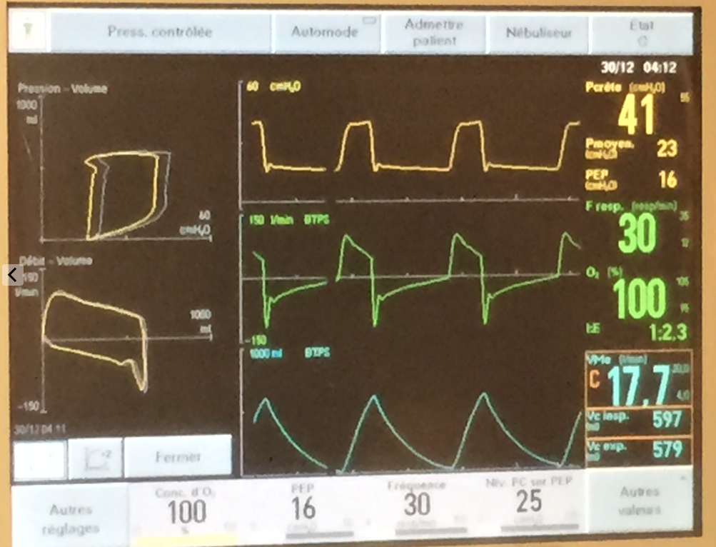
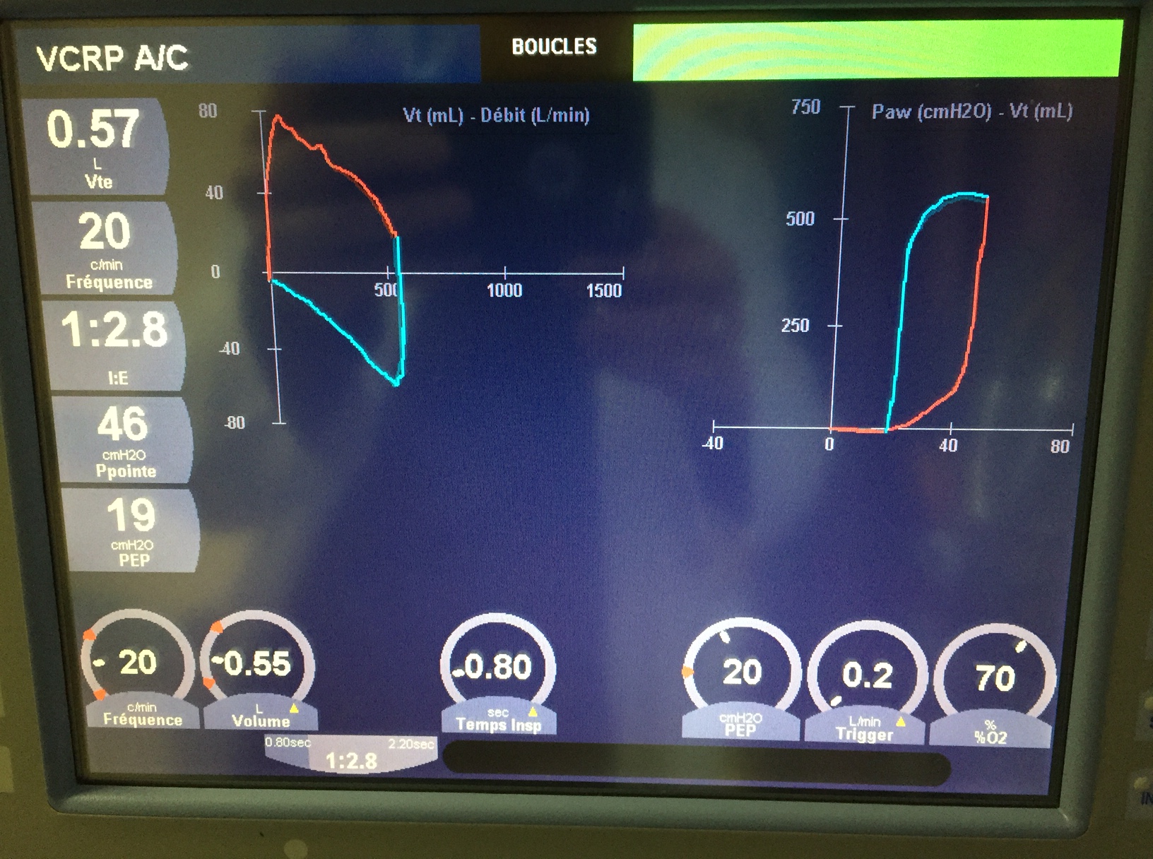
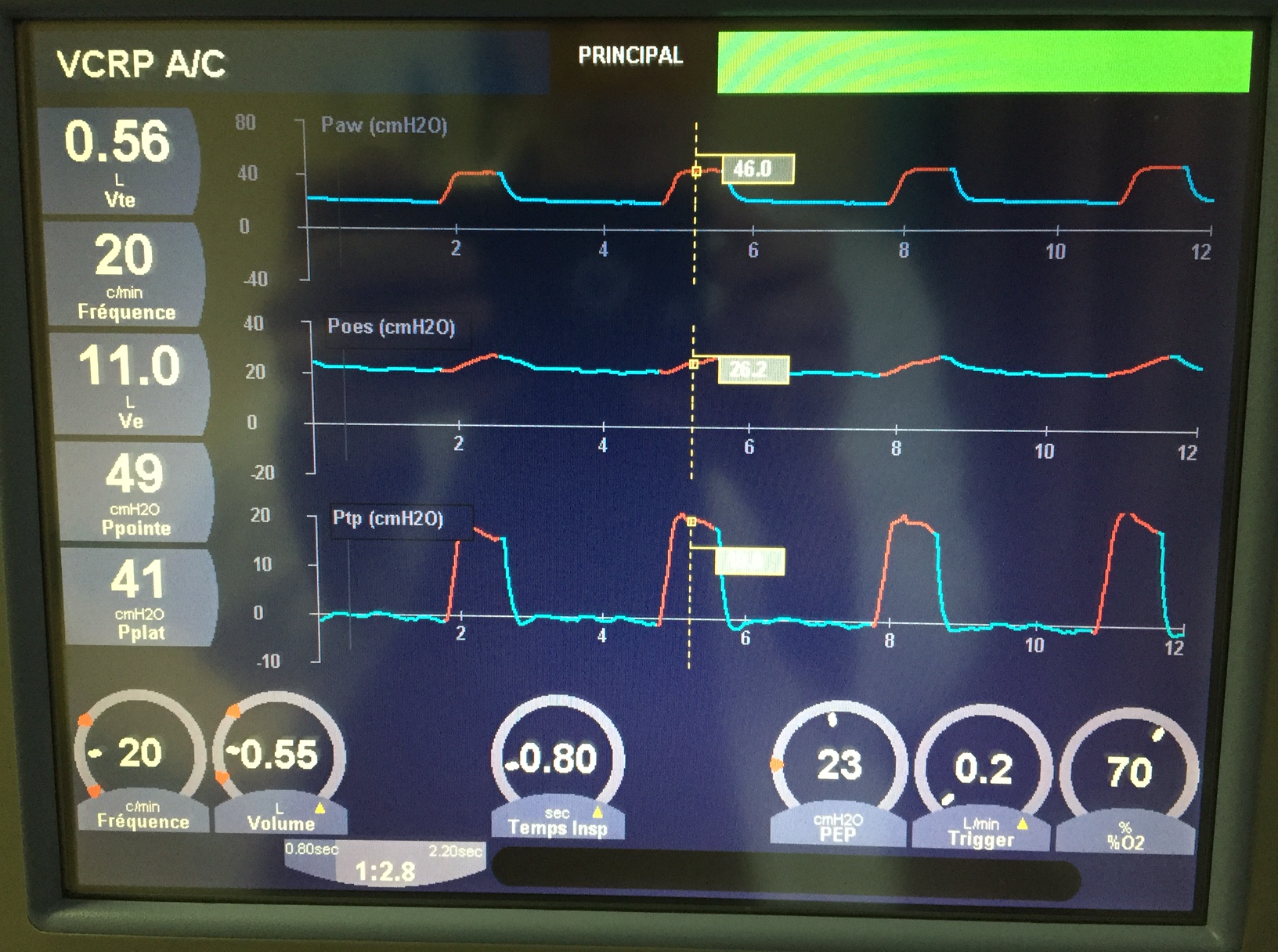
The case exposure will be mainly ICU as well as ER and ward patients. The focus will be on acute care issues. After two days, participants who had a basic ability in ultrasound should be fairly comfortable with assessing volume status, cardiac function, perform lung ultrasound, be able to identify and assess intrathoracic and intr-abdominal fluid collections, assess the kidneys, bladder and gall bladder, measure optic nerve sheath, assess carotid flow and some may have exposure to trans-cranial doppler. The focus may be shifted depending on a participant’s interest.

This takes place in Montreal, Quebec or Toronto, Ontario, Canada.

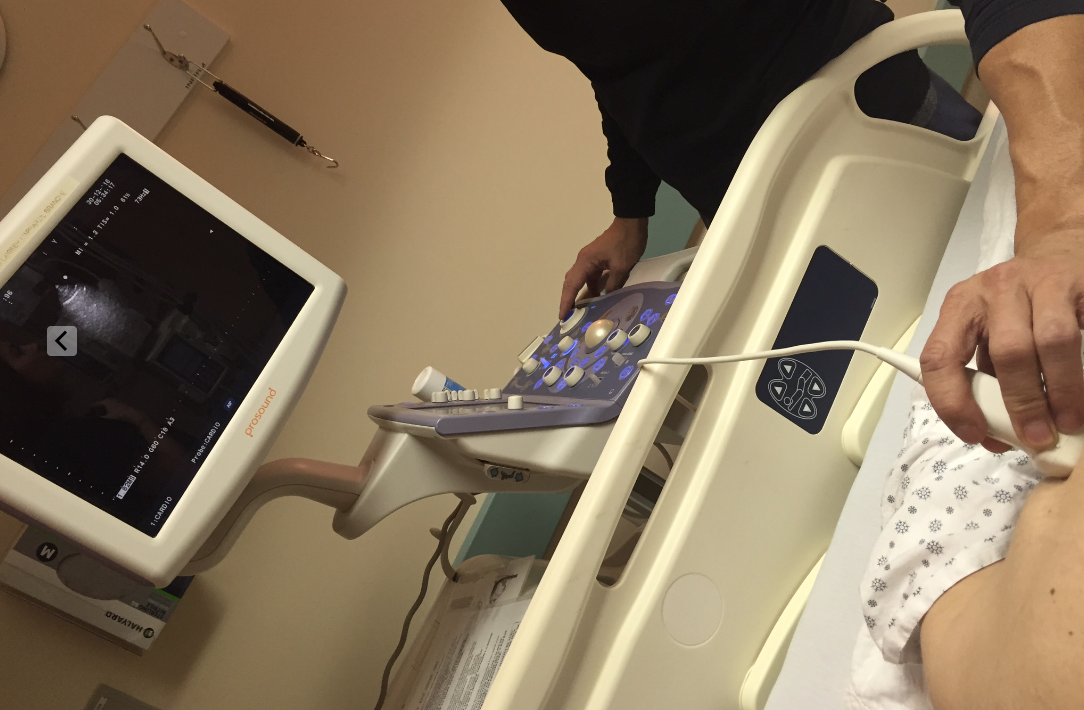
Participants will have the opportunity to work with handhelds, midrange and high-end ultrasound devices.

Space is limited as we can generally only accommodate 1-3 participants per month.

CME

So, great news, finally went thru the CME process and lo and behold, the Mini-Fellowships qualify for 25 Section 2 credits (regardless of the length) and 3 hours of Section 3 credits (per day of fellowship). For you americans:

Through an agreement between the Royal College of Physicians and Surgeons of Canada and the American Medical Association, physicians may convert Royal College MOC credits to AMA PRA Category 1 Credits™. 

Bonus!

Upcoming participants will also receive a copy of the forthcoming handbook:

covercover-back

Requirements 

Please have basic experience in bedside ultrasound. We don’t want to teach you about depth and gain. We’re happy to fine tune your views but not to introduce you to the main cardiac views. It would just be wasting your clinical time. We’re here to show you how to assess pathology and integrate your findings into clinical decision-making. Take the basic group course to learn the views, or be self-taught from youtube/iphone and practicing on your patients. You don’t have to be great, but to get the most out of this experience it shouldn’t be your first time holding a probe.

Registration

email me at philipperola@gmail.com or reach out on twitter @ThinkingCC

Tuition (Updated 2022)

Montreal Mini-Fellowships: 600$ CAN / 500$ USD per day for 1 physician, 400$ CAN / 350$ USD per person per day for additional days, and 400$ CAN / 350$ USD per person per day for a 2-3 physician group (maximum)

Toronto Mini-Fellowships: 800$ per half day (4h).

100% refundable until you start. Even if you don’t show up. Really. We’re not in it for the business. We get to go home earlier if you don’t come.

Testimonials:

« I have had the chance to participate in a shadowing experience with Dr Rola at the Scarborough General Hospital ICU during two days in 2013. As a general internist and assistant program director, this experience really opened my eyes regarding the use of bedside ultrasound in general internal medicine and for IM residents. I think I would have benefited more of this experience if I had done more training previously, and I encourage future participants to do so. However, I came back from this experience with a very clear idea of the benefit of CUSE for my patients and for our residency training program. I really saw how ultrasound was used ‘in action’, in a much more realistic way than what is usually shown in CPD meetings. I also saw its limitations and the skills I needed to develop to generate good images (not something you can learn over the weekend!). Since then, I participated in formal trainings and licensing activities (more than 250 supervised US on acute care patients) and now practice bedside ultrasound autonomously. We now offer a bedside ultrasound training for our residents with the help of the emergency medicine department and an ultrasound-guided procedural simulation lab. Nothing in CPD has improved my practice and benefited the health of my patients as much as bedside ultrasound training. »

Alexandre Lafleur, MD, MSc (Ed.), FRCPC
Spécialiste en médecine interne
CHU de Québec – CHUL
alexandre.lafleur.1@ulaval.ca

“Thank you very much for the exposure and teaching offered via the CCUS “Mini-Fellowship.”  These few days allowed me to enormously improve my mastery of bedside ultrasound in clinical decision-making in critical care. I recommend the experience to clinicians already having experience in bedside ultrasound, but who feel they could benefit from the expertise of an instructor to attain a level beyond basic courses and available textbooks.”

Mathieu Brunet, MD, GP/ER/ICU, Magdalen Islands, Quebec, Canada

“The CCUS Mini Fellowship In House training is very essential in to experience the echo skills that we get from the courses,being supervised in ICU will offer the chance to be corrected and get real live practice/exposure by being at the bedside and learn what is priority in echo for the best of patient care. The in-house experience is very helpful, practical, I recommend this training to any physician involved in ER, ICU, CCU, Anesthesia and rapid response team.”

Joe Choufani, MD, Internal Medicine/Cardiology, St-Lawrence Health Association, NY

“Thanks for everything. I really appreciate you sharing your vast fund of knowledge with me.”

Sean Sue, MD, ER, Philadelphia

CME

So, great news, finally went thru the CME process and lo and behold, the Mini-Fellowships qualify for 25 Section 2 credits (regardless of the length) and 3 hours of Section 3 credits (per day of fellowship). For you americans:

Through an agreement between the Royal College of Physicians and Surgeons of Canada and the American Medical Association, physicians may convert Royal College MOC credits to AMA PRA Category 1 Credits™., #CME