Wicked Clinical Case: POCUS & Prone save the day! #FOAMed, #FOAMcc, #FOAMer

So I get a call from a colleague in the ED at about 2am, telling me about a 39 yr old woman post-arrest. So I start putting on my boots and warming up the car (it’s January in Montreal folks).  Apparently she had presented earlier in severe acidosis, the diagnosis is unclear, but she apparently got 2 units for an Hb of 49, then went into respiratory failure and got intubated. She arrested about 30 minutes later, cause unknown.

I tell the ICU to prepare a bed but I want to see her in the ED first. Twenty minutes later I put probe to patient and see a full IVC with spontaneous echo contrast. On that I tell the nurse to hold the fluids – there was a bag and tubing and a pump with 100ml/hr on it – and turn into a subxiphoid view to see a normal RV and a hypokinetic LV with some WMAs. She has marked consolidations  in both posterior lung fields and B lines laterally, with small effusions and dynamic air bronchograms (indicating patent airways). At this point she has a HR of about 120, but there is neither perceptible BP (by NIBP) nor saturation. She’s on levophed at 20mcg. She’s about an hour post arrest which was witnessed and brief (<10min to ROSC).

The theories about the arrest are possible hyperkalemia: she was intubated with succinylcholine before the K of 6.1 was back from the lab, and her pre-intubation pH was 7.0, and post-intubation she was only ventilated at 400 x 18, possibly precipitating a drop in pH and a rise in K. Her EKG had some nonspecific signs at this point, but also a poor anterior R wave.

So we head to the ICU, as instrumentation was needed. Cerebral saturation (SctO2) is 42% and ETCO2 is 20mmhg, which reassures me that the BP is probably in the measurable range (normal SctO2 is >60% and varies, but 47% is certainly viable)…  A jugular CVC with continuous ScVo2 and a femoral arterial line goes in:

screen-shot-2017-01-05-at-10-44-50-pm

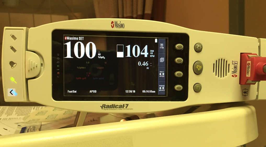
So with a BP of 59/44 (ignore the 100/46, not sure whose arm that was on!) I start epinephrine, as the POCUS is similar, as I want some added beta-agonism. ScVO2 matches SctO2 in the 40’s. We get the BP up the the 90-1oo range, the ETCO2 goes to 30, the SctO2 and ScVo2 go up into the high 40’s, which is very reassuring, because with this I know that my epi drip is improving perfusion and NOT over-vasoconstricting. Without looking at a real-time tissue perfusion index of some sort or other, it is nearly impossible to know rapidly whether your therapy is helping or harming (will discuss tissue saturation & resuscitation monitoring in more detail in another post sometime soon).

screen-shot-2017-01-05-at-10-46-31-pm

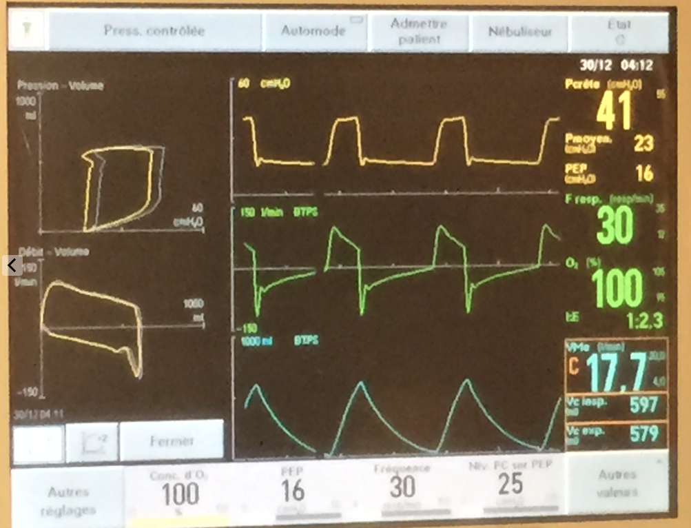
So now the sat finally starts to record in the low 60’s. We have a PEEP of 5, so start bringing it up. We hit 16 before the BP starts to drop, and that only gets us to the mid 70’s sat%. She actually squeezes my hand to command.

screen-shot-2017-01-05-at-10-45-21-pm

At this point I take a few seconds to recap in my mind. I’d spoken to the husband briefly and she had had recurrent episodes of feeling unwell with headache, nausea and diaphoresis, and that had been out for dinner earlier and she felt fine until later in the evening when this came on and eventually brought her to hospital. There was also a notion of hypertension at an ER visit a couple of weeks ago. Her history was otherwise not significant. Nonsmoker.

Pheo? Maybe, but shock?  I repeat the EKG, and now, in I and AVL, there is perhaps a 1mm ST elevation. She’s 39 and essentially dying. Lactate comes back >15, pH 6.9.  I give her a few more amps of NaHCO3. You can see the BP respond to each amp. I decide we need to go to the cath lab and get the cardiologist on call to get on the horn with the interventional team at a nearby hospital with a cath lab and ECMO, which is what I think she needs. Hb comes back at 116, making that initial 49 that prompted 2 PRBCs probably a technical or lab error…very unfortunate. There are no visible signs of significant bleeding.

But back to the patient, because this isn’t really a transferrable case.

Recap: a 39yr old woman in cardiogenic shock AND in severe congestive heart failure exacerbated by fluids and packed red cells, with a PO2 in the 40’s and sat in the 70’s.

So I decide to prone her.

screen-shot-2017-01-05-at-10-47-44-pm

Along with draining tamponades, this had to be one of the most rapid and rewarding maneuvers I’ve done. There was a scry drop of sat to the 40’s for a few seconds (may have been a technical thing), but then within a few minutes: BP to the 130’s, SctO2 to 59% and sat 100%!

screen-shot-2017-01-05-at-10-46-46-pmscreen-shot-2017-01-05-at-10-47-31-pm

screen-shot-2017-01-06-at-12-08-05-am


We dropped the vasopressors, the FiO2, and all breathed a collective sigh of relief. Now for the novices out there, prone ventilation improves VQ mismatch by moving perfusion from diseased, posterior lung fields to now-dependant, relatively healthy, anterior lung fields.

So transfer at this point was in the works. I planned to leave her prone until the last minute. The miraculous effect started to slowly wane within about 30 minutes, with sat and BP creeping down. At the time of transfer, we were back up to 80% FiO2.

So why is this?  Simple enough, this being simple pulmonary edema – rather than consolidated pneumonia – it migrated to dependent areas  relatively quickly. This was confirmed by a quick POCUS check:screen-shot-2017-01-05-at-10-48-06-pmscreen-shot-2017-01-05-at-10-48-26-pm

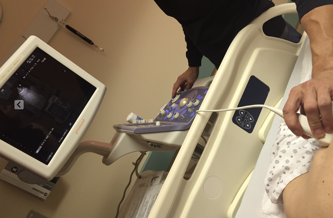
So in the still shots, you see a pristine “A” profile (normal, no edema) from the patient’s back, and a severe consolidation or “C” profile with ultrasound bronchograms in the antero-lateral (now dependant) chest. Impressive. (for those wanting some POCUS pearls see other posts and here). This is the reverse of her initial POCUS exam.

So we flipped her back and transported her – lights & sirens – the the cath lab, where they were waiting with ECMO cannulae. As an aside, it was quite refreshing to speak to the ICU fellow who spoke POCUS as well as french and english – it’s not usually the case, but I’m glad to see the change. I do believe it to be a direct effect of the influence of my friend and mentor, Dr. Andre Denault, one of the POCUS deities.

So she turned out to have a normal cath and a large adrenal mass. She did well on ECMO, being weaned off it today, and is now alpha-blocked and waiting for surgery, neurologically intact for all intents and purposes. A big thanks to the interventionists and the ICU team at the Montreal Heart Institute. Puts a smile on my face.


Take Home Points:

  1. don’t resuscitate without POCUS. I wouldn’t want anyone guessing with my life on the line, would you?
  2. keep pheo in mind as a cause of “acute MI” and shock
  3. if you’re not using some form of realtime monitor of perfusion (continuous CO, SctO2, ETCO2, ScvO2) then all you’ve got is looking at the skin and mentation, so you are essentially flying blind. Lactate and urine output are not realtime in real life.
  4. get ECMO in the house, it’ll come in handy. I’m working on it.


Love to hear some comments!

cheers


Philippe


ps I’ll try to add more ultrasound clips from this case in the next few days.

MOPOCUS: A great synopsis by Ha & Toh. #FOAMed, #FOAMcc, #FOAMus

Just came across this review and figured I should share. The authors make a great synopsis and review of POCUS in acute illness:

MOPOCUS Review by Ha &To

The only thing I would add to this is a more physiological way to assess the IVC, which I’ve blogged about here.  Sadly, I’ve heard a few people stating how they didn’t want to get into the dogma of IVC ultrasound, that it wasn’t reliable, etc.  The IVC doesn’t lie. It’s just not a recipe. The IVC findings have to be integrated into the rest of the echo graphic and clinical examination.  Trying to use it as a single value is akin to using serum Na+ as a diagnostic test for volume. It works only sometimes.

Please spread among the POCUS non-believers. We’ll convert them, slowly but surely. But the sooner, the better for the patients. Again, there’s no excuse to practice acute care without ultrasound. It’s not right. I’m not saying every probe-toting MD is better than one without, but everyone would up their game by adding POCUS, once past the learning curve!

cheers!


Philippe

Tom Woodcock: The Revised Starling Principle and The Glycocalyx! #FOAMed, #FOAMcc

Screen Shot 2016-08-05 at 11.57.11 PM

So today, I had the chance of having a private tutorial with Dr. Thomas Woodcock (@thomaswoodcock) about the glycocalyx and the revised Starling principles.  For anyone interested in fluid resuscitation, this is an area you have to delve into. The basic principles we all learned (which are still being taught) are basically the physiological equivalent of the stick man we all started drawing as toddlers: overly simplified and far from an accurate representation of reality.

Now my first disclaimer is that I have been a colloid supporter for many years. My physiological logic for that had been to minimize the crystalloid spillover into inflamed/septic areas, particularly the lungs and abdomen, when those are the septic sources. However, I was likely misled by my education and lack of knowledge about the endothelium.

So I stumbled upon the whole glycocalyx thing a couple years ago, and this prompted me to try more enteral fluids – the only way fluids normally ever enter the vasculature – but little else. Aware that it’s there, but unsure what to do about it.

Now a year and a half ago, Andre Denault, my closest thing to a mentor, casually dropped the line to me about albumin not working. “Don’t use it. It doesn’t act the way we think it does.”  But it was a brief chat, and I didn’t get to pick his brain about it.  Just a few weeks ago, I discuss with Jon Emile (Kenny), and he’s coming to the same conclusion.  Damn. I’m finding it a bit harder to hang on to my albumin use, which is beginning to look a bit dogmatic and religious.

Here is Jon-Emile’s take on it – a must-read.

Here is Tom Woodcock’s site and article – another must-read.

And here is my discussion (in two parts) with Tom (to skip the silence, skip forward to about 30 seconds into each – sorry my editing skills are limited!)


Bottom line?

Probably stick to isotonic crystalloids, and some hypertonics.


Love to hear some thoughts!

Cheers


Philippe


The NYC Tracks with Jon-Emile: The Glycocalyx – The Next Frontier. #FOAMed, #FOAMcc

I was really psyched when Jon-Emile mentioned he would like to talk about the glycocalyx.  I first blogged about it here, basically when I stumbled on the extensive literature on this huge organ we have been completely ignoring in terms of physiology and therapeutics. It lines our entire endothelium, which is where most of our therapeutic interventions go, and we only heard of it in passing, possibly in histology class as med 1’s.   Hmmm.  Anyhow, here, Jon-Emile and I talk about it a little, discuss possible clinical implications, but more importantly Jon mentions the relatively new blog of Dr. Thomas Woodcock (@thomaswoodcock), http://www.fluidtherapy.org, who is one of the pioneer clinicians who have studied the glycocalyx, and who is now trying to bridge the bench to the bedside.

I’ve been fortunate enough to get in touch with him and we’re planning to record some discussions soon.

So, in my view, the glycocalyx is a formidable force we have been ignoring, and have been damaging often with our interventions. I’m hoping to see some developments allowing glycocalyx assessment outside of the labs in order to give us the tools to reassess every fluid in terms of the relative damage it does to what is essentially the gatekeeper between the blood and the tissues.

Love to hear some comments!

Here is the chat with Jon:


cheers


Philippe

The NYC Tracks with Jon-Emile: Paracentesis and Volume Status. #FOAMed, #FOAMcc, #FOAMus

So I was in NYC last week and met up with my buddy Jon-Emile Kenny, (@heart_lung), intensivist-physiologist extraordinaire, and we recorded a few discussions on practical matters.

I always love to debunk myths and avoid dogmatic guesswork, and, more often than not, Jon, with his encyclopedic knowledge of the physiology literature, but more importantly a cutting edge understanding of it, can back up my vague ideas and empirically derived ideas, so that the next time someone asks me why this is so, I can have a semi-enlightened answer!

So here is the first, where we discuss the common question about the need (or not) of intravascular volume repletion during or following large volume paracentesis. Yes, there are some formulas out there as to how much albumin or crystalloid one should give, due to the worry of subsequent hypovolemia. Note how those formulas use no data about your patient’s volume status at the time of paracentesis, so as far as I’m concerned, they have no value whatsoever in an era where we can assess this. Yes, ultrasound is the base as far as I’m concerned.

Here we go:

Please share your thoughts!

cheers


Philippe

Fluids in Sepsis: An EmCrit Webinar! #FOAMed, #FOAMcc

Screen Shot 2016-04-27 at 2.00.28 PM

Screen Shot 2016-04-27 at 1.43.23 PM

So a few weeks ago Scott (@EmCrit) asked me to be part of a pretty cool webinar organized by the Greater New York Hospital Association about fluids in sepsis. The gang consisted of David Gaiesky, Emmanuel Rivers and moderated by Scott himself. And for some obscure reason, he asked me to be part of it – much to my honour (terror, also), naturally.  It was only afterwards that he told me it was to help stir the pot and be controversial, challenge the “old school” etc… He seemed to have overlooked that I am Canadian, and inherently and perhaps overly polite and considerate – at least live and in “person”!

We talk about a bunch of stuff around fluids, which, how much, how to assess, etc.

Anyhow, I hope I got a few ideas across, but it was really cool to hear that these gurus do use ultrasound – don’t necessarily strictly adhere to, for instance, EGDT, and also advocate that guidelines are guidelines and not necessarily gold standards.

Here is the link to the webinar for those interested:


https://t.co/dbL03Vuqlj


And here is the figure for the section where I refer to fluid responsiveness/tolerance:

Screen Shot 2016-02-21 at 9.25.50 AM

I further talk about this in a previous post here.

Scott and I also recorded a debrief which should be coming up in the next weeks on EmCrit – link to follow!

cheers!


Philippe

Cerebral & Somatic NIRS (Near InfraRed Spectroscopy) in shock states: tailoring therapy. (PART 1) #FOAMed, #FOAMcc

So I’d mentioned using NIRS to monitor and tailor therapy a few months ago, and promised a more in-depth discussion to come, so here we go.

For this not familiar with the technology or the concept, NIRS measures tissue saturation, predominantly venous. Hence physiologically it is akin to central/mixed venous gases, but localized. Cerebral NIRS found its foothold in the OR with carotid and cardiac surgery, but its use is now expanding. Given typical knowledge translation time of a decade, it should end up joining ETCO2 as a routine vital in monitored units, but probably not soon enough.

So in our unit at Santa Cabrini Hospital in Montreal, we’ve had this technology for about a year (the INVOS system), and have been studying its uses. In this time, three applications have stood out:

  1. Finding the “Sweet Spot” for vasopressors.
  2. Confirmation that therapeutic interventions are hemodynamically appropriate.
  3. Cardiac arrest: CPR adequacy, prognostication and detecting ROSC.


  1. Finding the “Sweet Spot” – I think (hope) that anyone reading this with professional interest understands that pressure does not necessarily equal perfusion.  With that in mind, adjusting vasopressors to a pressure makes little sense, and represents at best a guesstimate of perfusion, which is what we really are after. We can all agree, however, that a certain minimum pressure is required, but whether that is 65, 55 or 45 MAP no one can say for sure.  So the way I like to use it is to establish a baseline and watch the direction of the tissue saturation with vasopressor therapy. If the saturation begins to drop off, we may have reached a point at which excessive vasoconstriction is worsening tissue perfusion, and that inflexion point may represent the upper beneficial limit of the vasopressor – this may happen to be under 60 or 65 of MAP.  However, it is key to understand that this inflexion point is reflective of the current state of hemodynamics, such that a change in volume status or cardiac output, in one direction or the other, would likely change the position of this physiological point.  For example,  a volume depleted patient may reach a decreasing tissue saturation point at 55 MAP, but, once volume replete, may reach a higher MAP of 65 or above before a drop in saturation is seen.  Conversely, a patient whose best tissue saturations were around 65 MAP who suffers an MI and sudden drop in cardiac output may now see his perfusion compromised at that same MAP, which would now be achieved with a greater vasoconstriction, less cardiac output and consequently, poorer flow… I posted a case discussion which illustrates this.
  2. Confirmation that therapeutic interventions are hemodynamically appropriate – I feel this is really important. When a patient’s life is literally on the line, and knowing that our interventions are seldom without potential nefarious side effects, it is poor medicine to be introducing a therapy without having some form of monitoring – preferably multiple – that we are headed in the right direction, or at least not making things worse. Of course, we already do this – with BP, sat, lactate, CCO, ultrasound, ETCO2 – but I think using a realtime measure of tissue saturation adds to this. It is also my firm opinion that integrated, multimodality monitoring is necessary – at least until someone develops some form of mitochondrial monitoring which tells us that the cytoenergetics are sufficient to survive. Until then we are stuck with surrogate markers and many of them (e.g. lactate) are the result of complex processes that preclude them being a simple indicator of perfusion adequacy. For instance, when giving a fluid bolus/infusion – after having determined that the patient is likely fluid responsive AND tolerant – one should expect to see an increase in ETCO2 (other parameters being constant), an increase in CO, an increase in NIRS values. The absence of such response should make one reconsider the intervention, because without benefit, we are left only with side effects.

Here is a patient’s cerebral (top) and and somatic (thigh – bottom) and CO values. This patient had an RV infarct and was in shock.

IMG_7948IMG_7946


Following initiation of dobutamine, this is what occurred:

IMG_7951IMG_7949

Given that we cannot always predict the response to an inotrope – depending on the amount of recruitable myocardium, it is reassuring to see an improving trend. This enabled us to decrease the vasopressor dose significantly.

Note that, so far, and unless some good evidence comes out, I don’t use a goal value, and so far, I have not identified a value that is predictive of prognosis. However, downward trends usually bode very poorly. For instance, I had a severe chronic cardiomyopathy patient whose cerebral saturation was 15%!!!  But more surprisingly, she was awake, alert and hemodynamically stable. Adaptation.

Part 2 and the stuff on cardiac arrest coming soon!

Please, anyone using NIRS in shock, share your experience!


cheers


Philippe

Volume responsiveness and volume tolerance: a conceptual diagram. #FOAMed, #FOAMcc, #FOAMus

So I know I’ve belaboured the point about the difference between volume responsiveness (i.e. will there be significant increase in cardiac output with volume infusion) and volume tolerance (is the volume I am considering giving going to have nefarious consequences), because in my opinion, the focus has been – rightly so to some degree – to look for an accurate way of discerning responsive patients from non. Of course this is absolutely necessary, as one does not want to give volume if it will not have any benefit, but the too-common corollary to that is to automatically give volume to those who are responsive.  Here is an earlier post about this:

Fluid Responsiveness: Getting the right answer to the wrong question. #FOAMed, #FOAMcc, #FOAMus

So in discussing with a bright young colleague yesterday, Dr. St-Arnaud (@phil_star_sail), I realized that there may be a common conception that physiologically, the relationship between the two may be the following:

Screen Shot 2016-02-21 at 9.03.01 AM

This would mean that it is safe to give volume until a patient is no longer volume responsive, and even perhaps a bit more. Alternately, the two may be closer:

Screen Shot 2016-02-21 at 9.02.27 AM

This would mean that once can go just till the point where the patient is no longer volume responsive.

Either one of these scenarios would be awesome. That would mean that by using any of the flow or volume variation techniques, arterial or venous, we could pretty much remain safe.

However…

While the above may hold true for healthy subjects, I would contend that in sick people (which is who I tend to deal with, especially when resuscitating shock), that the more likely physiological relationship is the following:

Screen Shot 2016-02-21 at 9.03.28 AM

Hmmm… That would mean that assessing for volume responsiveness would only tell you that there would be an increase in cardiac output, but absolutely nothing about whether it would be safe to do so.

This concept is not a new one by any stretch of the imagination. It’s inferred in the diagnosis of “non-cardiogenic pulmonary oedema.” So what causes this shift? Here:

Screen Shot 2016-02-21 at 9.25.50 AM

So, how do we figure out where the point is? Sorry to say there is no answer that I know of. My friend Daniel Lichtenstein uses the FALLS Protocol (identifying the appearance of B lines during resuscitation) which is the least we should do, but I suspect that at that point, we have already overshot the mark. My adopted mentor Dr. Andre Denault (@Ad12andre, in addition to IVC, has identified portal vein characteristics including pulsatility (lots of stuff in press) to show that the viscera are at risk, but as of yet there is no simple answer. CVP value? Please. CVP tracing morphology? Maybe.

No simple answer. No one-size-fits-all velue to look for. Clinical integration.

In my opinion, one should not, in sick patients, seek to volume resuscitate until the point of no-volume-responsiveness. The old adage of “you have to swell to get well” likely kills a few additional patients along the way, just as much as under-resuscitation. I plead guilty for over-resuscitating patients for years before realizing that being on the flat part of Frank-Starling is 100% a pathological state.

Love to hear your ideas and comments!


Jon-Emile Kenny says:

I like your graphics, it makes the concepts tangible. I think we should try to integrate ‘volume status’ into this framework as well. A physiological purist might say that as soon as you are ‘hypervolemic’, you are volume intolerant, because hypervolemia is an abnormal state which should always be avoided. A functionalist might say that you become volume intolerant as soon as you have physiological embarrassment of any organ system – but how is this determined? My gut is that by the time there are B-lines in the lung, you’ve gone too far. By the time there is abnormality of splanchnic venous return, you’ve already gone too far. I am more of a purist, so in my perfect ICU, I would perform q4-6 hour radio-labeled albumin studies to determine the patient’s true plasma volume. In health, the normal blood volume is about 80 mL/kg [thus, once you’ve given a 70kg man 5 L of NS, you’ve almost certainly replenished his vascular volume]. The moment that the blood volume becomes > 95% the norm, I would call the patient volume intolerant and stop volume expansion and focus on venous tone with pressors, cardiac function with inotropes, etc. To me, this makes the most sense in the pure Guytonian world; if you keep flogging a patient with litre after litre of fluid and the patient’s BP remains low, you are missing something – volume is not the answer – regardless of what an ultrasound shows you:
1. trouble shoot the venous return curve [i.e. too little blood volume, too little venous tone, too high resistance to venous return]
2. trouble shoot the cardiac function [i.e. poor rate, rhythm, contractility, valve function, biventricular afterload]
If you need some objective measure of blood volume before you can call volume status optimized before moving onto the next problem to fix – that’s a radio-labeled albumin.
Maybe I’m crazy.cheers

Jon

Thanks for commenting Jon!

I totally agree, if we knew each patient’s normal blood volume, that would be a starting point.  And of course, that would prevent the over resuscitation of a very dilated and compliant venous system (small IVC on ultrasound). Let us know if you figure out a practical way to do that!

It’s too bad that extravascular lung water doesn’t seem to have panned out – not sure why exactly.


Philippe

Musings with Jon-Emile & Philippe – Fluid Resuscitation: Physiology and Philosophy! #FOAMed, #FOAMcc, #FOAMer

So here, Jon-Emile and I explore a topic I’ve posted about before (http://wp.me/p1avUV-bd) so I can see if a master physiologist agrees with my rationale (…not just my rationale but supported by a ton of literature many choose to overlook!).

Please visit http://www.heart-lung.org for Jon’s awesome physiology tutorials!

Love to hear listeners’ thoughts!

cheers


Philippe

Musings with Jon-Emile & Philippe 2: Putting Jon on the Spot!!! #FOAMed, #FOAMcc, #FOAMer

So in this second instalment, I put Jon-Emile on the spot about some common clinical scenarios which, to me, contain a bit of dogma. Let’s see if physiology will give us the bottom line!


I think these are actually really important, because just too many times, I hear people automatically saying that in RV infarct, the patients need a lot of fluids, and in PE and tamponade as well.  I’m not so sure that’s always true, so I thought it would be a good idea to review this physiology with s real pro!


enjoy, and love to hear some comments!


cheers


Philippe