Fluids and Vasopressors in Sepsis, Wechter et al, CCM Journal: Anything Useful? #FOAMed, #FOAMcc

A couple of articles on fluid resuscitation worth mentioning. Not necessarily for their quality, but because they will be quoted and used, and critical appraisal of the content and conclusion is, without a doubt, necessary to us soldiers in the trenches.

The first one, Interaction between fluids and vasoactive agents on mortality in septic shock: a multi-center, observational study, from the october issue of the CCM Journal (2014) by Wechter et al, for the Cooperative Antimicrobial Therapy of Septic Shock Database Research Group, is a large scale effort do shed some light on one of the finer points of resuscitation, which is when to initiate vasopressors in relation to fluids in the face of ongoing shock/hypotension.

So they reviewed 2,849 patients in septic shock between 1989 and 2007, trying to note the patterns of fluid and vasopressor therapy which were associated with the best survival.  They found that survival was best when combining an early fluid loading, with pressors started somewhere in the 1-6 hour range.  I do invite you to read it for yourself, it is quite a complex analysis with a lot of permutations.

So…is it a good study?  Insofar as a retrospective study on a highly heterogeneous bunch of patients, I think so. But can I take the conclusion and generalize it to the patient I have in front of me with septic shock? I don’t think so. In all fairness, in the full text conclusion the authors concede that this study, rather than a clinical game-changer, is more of a hypothesis generator and should prompt further study. That, I think, is the fair conclusion.

In the abstract, however, the conclusion is that aggressive fluid therapy should be done, withholding vasopressors until after the first hour.  This is somewhat of a concern to me, since it isn’t uncommon for some to just read that part…

So why is this not generalizable?  First of all, I think that the very concept of generalizing is flawed.  We do not treat a hundred or a thousand patients at a time, and should not be seeking a therapeutic approach that works best for most, but for the one patient we are treating. Unfortunately, this is the inherent weakness of any large RCT and even more so in meta-analyses, unless the right subgroups have been drawn up in the study design.

Let me explain.

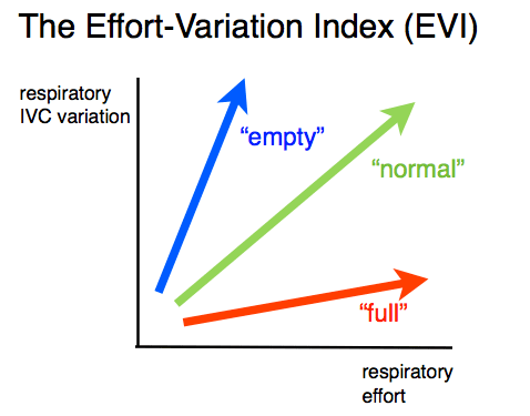
Patient A shows up with his septic peritonitis from his perforated cholecystitis. He’s a tough guy, been sick for days, obviously poor intake and finally crawls in. If you were to examine him properly, you’d have a hard time finding his tiny IVC, his heart would be hyperdynamic, his lungs would have clear A profiles, except maybe for a few B lines at the right base. You’d give him your version of EGDT, and he’d do pretty well. A lot better than if you loaded him with vasopressors early and worsened his perfusion. Score one for the guideline therapy.

Patient B shows up with his septic pneumonia, also a tough guy, but happens to be a diabetic with a past MI. He comes is pretty quick cuz he’s short of breath.  If you examine him properly, he has a big IVC, small pleural effusions, right basal consolidation and B lines in good quantity. He gets “EGDT” with an aggressive volume load and progressively goes into respiratory failure, which is ascribed to his severe pneumonia/ARDS, but more likely represents volume overload, as he was perhaps a little volume responsive, but not volume tolerant. An example of Paul Marik’s “salt water drowning.” (http://wp.me/p1avUV-aD) Additionally he goes into acute renal failure, ascribed to severe sepsis, but certainly not helped by the venous congestion (http://wp.me/p1avUV-2J). If he doesn’t make it, the thought process will likely be that he was just so sick, but that he got “gold standard” care. Or did he?

It may very well be that the studied group may include more Patient A types, and less B types, whose worse outcome will be hidden by the “saves” of the As. If you have a therapy that saves 15/100 but kills 5/100 you still come out 10/100 ahead… Great for those 15, not so much for the 5 outliers.

We, however, as physicians, need to apply the N=1 principle as we do not treat a hundred or a thousand patients at a time. I would not hesitate to be much more conservative in fluid resuscitating a B-type patient, regardless of the evidence.

Unfortunately, until trials include a huge number of important variables (an accurate measure of volume status, cardiac function, capillary leak, extravascular lung water, etc), it will be impossible to extrapolate results  to an individual patient.  These trials will, I suppose, eventually be done, but will be huge undertakings, and I do look forward to those results.

So, bottom line?

It’s as good a study of this type as could be done, but the inherent limitations make it of little clinical use, unless your current practice is really extreme on fluids or pressors. What it will hopefully be, however, is an onus to do the highly complex and integrative trials that need to be done to determine the right way to treat each patient we face.


thanks!


Philippe


COMMENTS:

Lawrence Lynn says:

Excellent post. This thoughtful quote should be read and understood by every sepsis trialists!!

“We do not treat a hundred or a thousand patients at a time, and should not be seeking a therapeutic approach that works best for most, but for the one patient we are treating.”

This single quote exposes the delay in progress caused by the ubiquitous oversimplification which defines present sepsis clinical trials. Bacteria (and viruses) generate “extended phenotypes” which are manifested in the host. These phenotypes combine with the phenotypic host response to produce the range of “dynamic relational hybrid phenotypes of bacterial and viral infection”. These hybrid phenotypes are also affected by the innoculum and/or the site of infection (vis-à-vis, your example of peritonitis).

Certainly Wechter et al and the Cooperative Antimicrobial Therapy of Septic Shock Database Research Group should be commended for beginning the process of moving toward the study of the dynamic relational patterns of complex rapidly evolving disease and treatment.

We are excited to see the beginning of the move of trialists toward the study of dynamic state of disease and treatment. However, before they can help us with meaningful results, trialists will need to study and define the range of “the dynamic relational phenotypes of severe infection” and then study the treatment actual phenotypes. This will not be easy as these organisms have had hundreds of thousands of years of evolution writing the complex genotypes which code for the extended of human infection. Sepsis trailists need to be encouraged by clinicians to rise to the task.

The clinicians must actively teach the trialists, (as you have in your post) that we expect trails which help to identity the therapeutic approach that works best in response to the dynamic hybrid phenotype “we are treating”.

The two linked articles below explain the present oversimplified state of the science of sepsis trails and why we clinicians must teach the trailists not to oversimplify and assure that they move quickly toward the study of the actual dynamic phenotypes of severe infection.

http://www.ncbi.nlm.nih.gov/pubmed/24834126

http://www.ncbi.nlm.nih.gov/pubmed/24383420

This is a paradigm shift so we, as clincians, must act to teach trailists this move is necessary. Otherwise we will continue to be left with hypotheses, which, while nice, are not useful at the bedside.

Lawrence Lynn


Another plea. Please stop embarassing us. #FOAMed, #FOAMcc.

Despite physiological rationale, common sense, and a JAMA article now almost 2 years old, I still sadly see most of my internal medicine colleagues still routinely reaching for (ab)normal saline.

Its embarrassing.

I genuinely feel bad recommending other fluids in consultations, or in the room of a crashing patient asking the nurse to stop the bolus of NS and change it at least to RL, because it is such a ‘basic’ intervention. Prior to the JAMA article, I mostly gave people the benefit of the doubt. Resuscitation isn’t everyone’s field of interest, nor is physiology, so I didn’t feel that necessarily everyone HAD to know this and ascribe to it. I do understand the 10 year time of knowledge translation, but that’s why #FOAMed exists, to try to cut that down.

So please, unless your goal is specifically chloride repletion, take a deep breath and release your grasp on habit and tradition, and embrace physiology (at least to some degree) and stop using NS as a volume expander whether in bolus or in infusion. RL or plasmalyte – although not physiological, at least not as biochemically disturbing as is 0.9% NaCl.

Having said that, let’s keep in mind that human fluid is colloid, whether it includes a cellular suspension (blood, lymph) or not (interstitial fluid), made of a varying mix of proteins, electrolytes, hormones and everything else we know – and some we don’t – floating around. There is no compartment that contains a crystalloid solution.

I’m quite aware that no meta-analysis has shown that colloids are superior, but it likely is just a matter of the right colloid. Resuscitating with crystalloids is kinda like throwing a bucketful of water at an empty bucket across the room. 70-80% spill, if you’re lucky. And the cleanup may be more costly than a few sweeps of the mop. This is evidence based (SOAP, VASST, etc..).

So a plea to all, spread the word. Its a simple switch. Boycott hyperchloremic acidosis at least.

For more details, here’s a link to my earlier post on NS: http://wp.me/p1avUV-5x

cheers


Philippe

CCUS 2014 – all lectures coming soon! #FOAMed, #FOAMcc, #FOAMus

The 7th edition of our symposium was awesome!  We had over 130 participants from all over Canada, the USA, and even had a few europeans hop the puddle and come share in the learning and practice!  All in all, two great days.


Thanks to the entire faculty, including #FOAMites Haney – @criticalcarenow, Mike – @bedsidesono, Vicki – @nobleultrasound, Taylor – @canibagthat, Jean Francois and Maxime – @EGLS_JFandMax who really made sure each participant had a great learning experience.


In the spirit of #FOAMed, we’ll be putting up ALL the lectures on the website in the next few weeks (www.ccusinstitute.org), and eventually a system for CME might be put in place. So make sure to bookmark or come back visit, as there were some really great lectures. Select workshops will also be uploaded.


Next year is in the works, so stay tuned!


Philippe

The Effort-Variation Index – a conceptual tool for IVC ultrasound. #FOAMed, #FOAMcc, #FOAMus

I recently had a colleague ask me to put on a graph the way I like to assess the IVC, at least conceptually.  I posted about this a few weeks ago (http://wp.me/p1avUV-8E), so I tried to come up with something useful for clinicians, correlating IVC variation with respiratory effort.

A useful concept to visualize this is the Effort-Variation Index (EVI). To obtain this, start by looking at a Frank-Starling curve, and broadly categorizing patients as being on the “empty” side, the “normal” or the “full” side.

Frank-Starling:Physiological


Next, if you look at how these groups would plot on a graph correlating IVC variation to respiratory effort, which, physiologically, would be the change in pleural pressure (delta Ppl), you should conceptually see something like this:


EVI


Note that this has not been validated, nor does it contain any values. It is simply, for now, a useful mental construct to understand the physiology behind the variability, and is useful when elaborating each patient’s physiological profile in the mind of the bedside clinician. Along any horizontal line, the IVC variation would be the same. You can therefore see that, given enough respiratory effort, a “full” patient could appear “normal” or even “empty.” Hence interpreting IVC variation without understanding this would lead to potential error.


Love to hear some thoughts and comments!


Philippe

Another wicked ultrasound case! Can you see the culprit? Another reason to do bedside ultrasound… #FOAMed, #FOAMcc, #FOAMus

Reviewing some TEE cases with Max Meineiri of TGH yesterday (Max is an anaesthetist-intensivist-sonographer extraordinaire who has been kind enough to help me brush up my TEE skills recently), here is one that stood out for two reasons. Here is the story: An 84 year old woman is sent from a peripheral hospital to the cath lab for chest pain.  She arrests on the table after they found normal coronaries and the code blue is called. Max arrives on the scene, and due to CPR making TTE difficult (and also because Max walks around with a TEE probe in a hip holster by Dolce & Gabbana), in goes the TEE probe and right away they note a massively dilated and hypokinetic RV, and a small and under filled LV. Yup, sure looks like a PE in these circumstances. Not being satisfied with a presumptive diagnosis, Max gets to a short axis view of the aortic valve and pulls out the probe slightly, following the bifurcation of the main PA.  On the screen, the right PA is on the upper left field, and the left PA disappears towards the upper right (the left main stem bronchus makes it difficult to visualize).

Anything seem a little odd?   Yup, you can see the occlusive culprit a couple of centimetres into the right PA, moving with each beat.  Being in angio already, they threaded a PA cath and administered thrombolysis, but despite some visual fragmentation, she did not survive. So why is this case interesting? 1. the image is pretty cool. 2. More importantly, it highlights the importance of bedside ultrasound.  If a rapid, focused cardiac exam had been done at her presentation at the peripheral hospital, the first-line physicians most likely would have noted the severe RV dysfunction and questioned the diagnosis of coronary syndrome, possibly (hopefully) thrombolysing the patient, and very possibly averting the cardiac arrest. …I know, I know, we don’t have all the info, the ECGs, etc, and maybe this was really an ACS and she happened to have a DVT which embolized during transport, etc…do you buy that?  Ockham and his parsimonious razor don’t, and I would tend to side with them.   love to hear some thoughts!   Philippe

Bedside Ultrasound: Quite a Case! #FOAMed, #FOAMcc

So here is an awesome clip from an ICU colleague of mine, Lorraine Law.  She was managing a post arrest (elderly woman who collapsed at home and was resuscitated but remaining in profound shock) case using bedside ultrasound and came across this pathology:

video courtesy of Lorraine Law & Shirish Shantidatt

what do you think?

scroll below for my thoughts…


So the clip starts with a subxiphoid 4 chamber view that clearly shows a massively dilated RV with a hyperdynamic and underfilled LV.

[For the hemodynamic novices, remember that the ventricles are kind of like roommates who share a pericardium. Especially in acute scenarios, if one gets overloaded, the other will have to give way, until the pressure equilibrates. If the process is exceedingly slow, they can do some renovations and stretch the pericardium, but this takes likely weeks. In this case, the elevated PAP overloads the RV and the RVDP > LVDP, resulting in decreased diastolic filling, which in turn drops the stroke volume/cardiac output/MAP.]

We can see that the RV TAPSE (tricuspid valve excursion towards apex) is really minimal, supporting an acute or acute on chronic process.

The clip then shows a long axis view of the IVC with echogenic material, most likely thrombus, with a to and fro motion, going in and out of the RA. Wow. You don’t see this very often.  The only thing preventing further travel is actually the fact that the cardiac output is so low due to massive embolism so that the flow can in fact barely carry the clots forward anymore at this point, similar to the sluggish IVC clip I put up a few months ago (http://wp.me/p1avUV-5t).

The most likely diagnosis is pulmonary embolism, and thrombolysis is indicated. Unfortunately despite my colleague’s timely diagnosis, the clot burden was likely too much, and despite thrombolysis, the patient passed away of intractable shock.  One can imagine that the TPA actually has to make it to the lungs, and with such a degree of obstruction, it is likely that very little actually got to the pulmonary vasculature…

Unfortunate case, but quite impressive images.

A crazy thought, using hindsight and with the luxury of knowing the fatal prognosis: intracardiac (RV) TPA bolus? Small spinal needle?  Anyone bold enough? Food for thought if (when) I see one like this…


cheers!


Comments:

Marco says:

Really quite impressive images. A couple of weeks ago I admitted a pretty young patient after a successful resuscitation due to massive pulmonary embolism. Immediately after ROSC in emergency department, he was transported to the cath-lab where TPA bolus was administered directly through a PA cathether. In ICU we continued the infusion. In less than 24 hours we obtained a relative hemodynamic stability and discontinued all the vasopressors, but the case remains unfortunate because despite therapeutic hypothermia the post-anoxic damage was so severe that led to cerebral death declaration two days later.


Thanks Marco, very interesting.  There is a recent study on catheter directed thrombolysis in PE reviewed at PulmCCM:(http://pulmccm.org/main/2014/randomized-controlled-trials/catheter-directed-thrombolysis-submassive-pe-better-heparin-rct/)

A physiological point about PE resuscitation is the relative inefficiency of CPR, as both venous return and LV filling is severely limited, so systemic perfusion is even worse than the usually poor output during chest compressions…

Thanks for reading!

Marco replies:

Thanks, Philippe!
The point about the possible inefficiency of CPR is crucial in my opinion. The patient I brought as example had a witnessed cardiac arrest (he called EMS when in respiratory distress) and CPR without interruption from the beginning, nevertheless he resulted in brain death declaration.
I remember very clearly a 43-year-old woman that 3 years ago had a massive PE in the OR shortly after a long lumbar vertebral stabilization. We admitted her to ICU after more than 80 minutes of CPR, a bolus of rTPA and with severe hemodynamic instability. RV was extremely dilated. When she eventually regained stability I had little hope about her neurological recovery, but surprisingly she was extubated the following day and last year she returned to our 12-months post-ICU follow-up showing perfect recovery.
I think that systemic and cerebral perfusion during “obstructive” cardiac arrests such as massive PE is very difficult to asses with current technology. A couple of times I was tempted to check it with trans cranial doppler, but usually there’s too much confusion during CPR.
When I was a resident I witnessed to a iatrogenic cardiac arrest in a patient with advanced monitoring that led to an interesting publication: http://www.researchgate.net/publication/10832333_Cerebral_perfusion_pressure_and_cerebral_tissue_oxygen_tension_in_a_patient_during_cardiopulmonary_resuscitation


Wow, very interesting cases.  What fortune to have been able to record that data, as obviously getting that in during CPR would be almost impossible.  TCD, at least after ROSC, could be contributory… Another option is using NIRS, which I’ll be working with this summer.

thanks again!

Philippe

An Update on Pulmonary Embolism: NEJM’s PIETHO Study…what’s the verdict? #FOAMed, #FOAMcc

As has been discussed in a previous post (http://wp.me/p1avUV-7T), patients with sub-massive PE (hypoxic, tachycardic, some troponin rise, etc…but no hypotension) remain in a grey zone, which is, to me , a dubious situation at best – their mortality can be up to 15%, morbidity likely more.  Everyone agrees the low-risk patients don’t need thrombolysis, and everyone pretty much agrees that the patient in shock needs it.  There is data out there suggesting that some patients clearly benefit from thrombolysis despite not being in shock, in good part relating to avoiding chronic pulmonary hypertension and its consequences.

The issue for many clinicians is that they have a “stable” patient in front of them, and they are considering giving them a drug that can potentially give them a bleed in the head and leave them dead or crippled. Many shy away from this. Part of this is cultural, because the same docs probably wouldn’t hesitate giving the drug to a lateral or posterior MI, which is not likely to kill you, or even leave you a cardiac cripple (just to be clear, I’m not advocating against thrombolysis in these cases, just trying to find a parallel), but since the AHA guidelines say to do it and everyone else does it, there’s no trepidation. It is the standard of care.  For most of us acute care clinicians who do not do outpatient medicine, if the patient survives and gets discharged home, chalk one up in the win column. But, as has become clear in recent years with the post-critical illness syndromes, morbidity can be just as important as mortality, especially in the younger patients. Kline et al (Chest, 2009) showed how almost 50% of “submassive PE” patients treated with anticoagulation alone had dyspnea or exercise intolerance at 6 months. They only had a 15% improvement in their pulmonary artery pressures (mean 45 mmhg).

What are the real risks? Pooling the data together gives a value around 2% with a spread between 0.8% and 8%, more or less. This represents each patient’s inherent risk of bleeding, as well as some of the inconsistencies with post-thrombolysis anticoagulation (safest to aim for 1.5-2 x PTT baseline in the first 48h).

The MOPETT trial which, as a #FOAMite you have certainly come across, showed that a half-dose of TPA was highly effective, and they felt it might be possible to go lower. The physiological beauty in that is that, unlike other sites we thrombolyse with full dose TPA, the lungs get 100% of the TPA (coronary artery gets maybe 5%, brain gets 15%).  Mind you, of course, the culprit clot/artery obviously doesn’t get 100%, but much, much more (if we figure that you need about 50% vascular area occlusion to cause RV dysfunction) TPA per “clot” than other pathologies. One can argue that anatomically, there is a greater clot burden than coronary or arterial thrombolysis, which may offset this somewhat. However, the date was quite clear in this trial that the therapy was effective, and the bleeding was none.

Ok, so let’s get to the PIETHO. 1000 patients, TPA+heparin vs heparin alone in normotensive but intermediate risk patients. So, first question is how was that risk defined?  Patients needed to have echocardiographic/CT signs of RV dysfunction AND a positive troponin. Interestingly enough, onset of symptoms was up to 15 days before randomization…not exactly early treatment, and unfortunately there is no information about the actual time to thrombolysis or subgrouping.  The results were as one could imagine. The combined endpoint of death or hemodynamic decompensation was 2.6% in the thrombolytic group vs 5.6% in the anticoagulation.  I’m not a fan of combined endpoints. Hemorrhagic stroke was 2.0% vs 0.2%. Their conclusion? Exercise caution. Hmmm…not much of a step forward. Basically tells us what we know. It helps the hemodynamics, but you can bleed. They do re-affirm that bleeding is more likely in the over-75.


What do we REALLY need to figure out? 

1. echographic risk stratification – at least into moderate and severe RV dysfunction.

2. longer term outcomes (hopefully PIETHO has a follow-up study in the pipeline, since they had good numbers).

3. a point-of-care study – time is of the essence, and may have an impact on dosage. IMHO thrombolysis should be done within a few hours of presentation at most.

4. further dosage data – 1/2? 1/3? 1/4? small boluses q1h until RV function improves?

I wish I could do it, but community hospitals don’t have the ideal setup, nor do I have a research team that can handle something of this scale. But surely someone can!


Bottom line?

It won’t change my practice. I will continue to offer thrombolysis in select cases, especially the younger patients, who obviously have a lower risk of bleeding, and stand to benefit the most, as pulmonary hypertension  can be crippling. I know I’d take the risk of bleeding when I see 50% dyspnea/exercise intolerance two years down the road…

Finally, bedside ultrasound to anyone with dyspnea/hypoxia should be a standard of care for every acute care physician. No ifs, ands or buts, no exception. Waiting for a CT angio or formal (read daytime hours) echocardiogram is, to me, unacceptable. If you, a friend or family member were in that ER bed, would you trust a physical examination and a CXR to rule out the need for an immediate intervention? I wouldn’t, not my own, and not even Dr. Bates’, Dr. DeGowin’s or Dr. Sapira’s, or all three combined.

cheers!


Kline JA, Steuerwald MT, Marchick MR, Hernandez-Nino J, Rose GA. Prospective evaluation of right ventricular function and functional status 6 months after acute submassive pulmonary embolism: frequency of persistent or subsequent elevation in estimated pulmonary artery pressure. Chest 2009;136:1202e1210.

Guy Meyer, M.D., Eric Vicaut, M.D., Thierry Danays, M.D., Giancarlo Agnelli, M.D., Cecilia Becattini, M.D., Jan Beyer-Westendorf, M.D., Erich Bluhmki, M.D., Ph.D., Helene Bouvaist, M.D., Benjamin Brenner, M.D., Francis Couturaud, M.D., Ph.D., Claudia Dellas, M.D., Klaus Empen, M.D., Ana Franca, M.D., Nazzareno Galiè, M.D., Annette Geibel, M.D., Samuel Z. Goldhaber, M.D., David Jimenez, M.D., Ph.D., Matija Kozak, M.D., Christian Kupatt, M.D., Nils Kucher, M.D., Irene M. Lang, M.D., Mareike Lankeit, M.D., Nicolas Meneveau, M.D., Ph.D., Gerard Pacouret, M.D., Massimiliano Palazzini, M.D., Antoniu Petris, M.D., Ph.D., Piotr Pruszczyk, M.D., Matteo Rugolotto, M.D., Aldo Salvi, M.D., Sebastian Schellong, M.D., Mustapha Sebbane, M.D., Bozena Sobkowicz, M.D., Branislav S. Stefanovic, M.D., Ph.D., Holger Thiele, M.D., Adam Torbicki, M.D., Franck Verschuren, M.D., Ph.D., and Stavros V. Konstantinides, M.D., for the PEITHO Investigators*, Fibrinolysis for Patients with Intermediate- Risk Pulmonary Embolism, N Engl J Med 2014;370:1402-11.

Mohsen Sharifi, MDa,b,*, Curt Bay, PhDb, Laura Skrocki, DOa, Farnoosh Rahimi, MDa, and Mahshid Mehdipour, DMDa,b, “MOPETT” Investigators, Moderate Pulmonary Embolism Treated With Thrombolysis (from the “MOPETT” Trial), Am J Cardiol 2012

Bedside Ultrasound Clip Quiz #8. #FOAMed, #FOAMcc

So a patient presents with worsening peripheral edema and right upper quadrant discomfort. Biochemical data only reveals some mild elevation of the transaminases.

Here is what you see:

These are the right sided chambers.

And this is a hepatic vein.


What do you think is going on?  Scroll below for the answer!


This patient has severe tricuspid regurgitation. The second clip shows significant retrograde flow in the hepatic vein (red doppler signal). Hepatic congestion was the cause of pain.

Analysis of hepatic vein flow can be useful to establish the presence of right heart dysfunction when cardiac views are difficult.


cheers!


Philippe

The IVC Assessment by bedside ultrasound: Let’s apply some common sense! #FOAMed, #FOAMcc

So I have a huge issue the IVC and its ultrasound assessment. For the most part, neither the yay-sayers or the nay-sayers are applying much sound physical principles, as far as I’m concerned.

To assess a patient’s volume status, it may be practical to begin with the sub-xiphoid view of the IVC, since the decision to give fluids or not – especially in emergency situations – can then be taken within the first few seconds of examining the patient. The physiological rationale behind assessing the IVC as a marker for volume responsiveness is simple and solid. As the venous compartment fills, the size of the IVC will gradually increase until it reaches a maximal size of about 20-25mm or even 30mm, depending on physical size and chronicity.

Concomitantly, the phasic respiratory variation will decrease as the venous pressure increases and the effect of varying intrathoracic pressure is no longer felt. At this point, the flat part of the Starling curve of the right ventricle is approaching, and there is little response to volume, and little physiological rationale to support giving more.

Currently, many use a variation of about 20% or more to suggest a volume responsive state in ventilated patients or an inspiratory collapse “sniff test” of 50% or more in spontaneously breathing patients.

There remains controversy around using IVC assessment for volume responsiveness, and with good reason! There are a few important reasons why:

a. technique – First of all, there is the manner in which IVC measurement has been taught: the M-mode measurement of the antero-posterior (AP) diameter during breathing (usually of ventilated patients) about 3 cm below the diaphragm. Although highly practical and reproducible, it has many shortcomings. If we look at the physiology, what changes with cycles of respiration is the volume of blood entering the chest/right atrium. Hence the key variable we are trying to assess is the transient variation in IVC size – which is a volume, not a linear dimension (I’m getting painful flashbacks of using pressure  to determine volume!)Hence the use of a single linear measure on one point along the length of the IVC to assess this is inherently flawed. For instance, the figure to the left shows how, if this IVC were to be measured in its AP diameter, the variability may not be that great. In the short axis, however, one can clearly see the significant change in surface area, and hence volume between phases of mechanical ventilation. Also, the IVC is rarely perfectly circular, but often ovoid, and occasionally with the greatest diameter in an anteroposterior axis, making that single AP measurement even less relevant.

b. intrathoracic pressure – Secondly, the variation in intrathoracic or intrapleural pressure (Pip) must be measured, as, for instance, a young and fit patient can generate large changes, which would result in more significant IVC variation, as compared to a frail elderly patient, even if they are on the same point of their Starling curve, invalidating the IVC measurement. All “sniffs” are not created equal.

c. intra-abdominal pressure – Finally, the intra-abdominal pressure (IAP) must also be assessed, since an elevated pressure would decrease the size of the IVC and make that measure no less accurate, but less relevant in terms of representing venous filling.

So what should we do?

Instead, a more global assessment of the IVC volume, measuring short axis area measurements and variation at several points along the IVC would give a much more accurate estimate of IVC volume variation. This is currently being studied by our group.

“Eyeballing the IVC” Attempting to link evidence and physiology, some bedside sonographers’ approach is to take a global look at the IVC in both long and short axis during respiration, while clinically assessing the respiratory effort and the abdominal pressure. This approach is analogous to the “eyeballing” of LV function – versus more formal measurements such as Simpson’s disk method, etc – which has been proven just as accurate with sufficient clinical experience.

The figure below shows an IVC that is about 10-12mm along most of its intrahepatic segment on expiration, and collapses almost completely on inspiration. If this belongs to a patient breathing with little effort and with a soft abdomen to palpation, it is physiologically quite clear that this patient would be fluid responsive. It also shows how impressive the collapse is in the short axis.

IVC insp dual

For instance, let’s say Patient A is in respiratory distress and using accessory muscles and presents to the ER with a respiratory rate of 35 and a systolic BP of 80. His IVC measures approximately 21 mm in diameter at several points along its axis, and has a brief collapse to about 10 mm with strong inspiratory efforts. His abdomen is soft during inspiration but firm during a prolonged expiratory phase.

Patient B is brought to the ER somnolent with a respiratory rate of 10 and a systolic BP of 80. His IVC measures 15 mm with minimal respiratory variation. His respirations are shallow and his abdomen soft. These two patients show how the IVC assessment needs to be taken in clinical context.

Patient A has some 50% inspiratory collapse of a large IVC in the context of large variations in Pip, whereas Patient B has little variation of a mid-sized IVC in the context of very small variations in Pip. In all likelihood, Patient A is not very volume responsive, while Patient B probably is.

Volume Responsiveness vs. Volume Tolerance – it is critically important to distinguish the difference between these two concepts as they are often misused interchangeably: Volume responsiveness refers to an increase in cardiac output (CO) to a fluid challenge. This is a purely hemodynamic concept. Volume Tolerance refers to whether or not a patient can tolerate volume without clinically significant side effects. This is a complex clinical assessment that should include: -the patient’s plasma oncotic pressure (serum albumin) and level of capillary leak, if present, -the patient’s pathology – is there risk of capillary leak in critical tissues such as lung, brain, abdomen? -the type of fluid being considered (isotonic crystalloid vs hypertonics or colloids/blood products). It is important to distinguish that not all patients who are volume responsive are necessarily volume tolerant.

Volume assessment summary – this issue remains a difficult one, even with the use of bedside ultrasound, because the optimal point for any one patient to be on his or her Starling curve at any one time in different clinical conditions remains elusive. Despite considerable study and several proposed management algorithms, there is no means by which to determine exactly how much of any given fluid is enough, without being too much.

In my opinion… – no direct evidence – keeping an IVC below 20mm and probably below 15mm with significant respiratory variation, if hemodynamics allow, is probably ideal. However, bedside ultrasound allows to clearly identify the cases where fluid is clearly needed and those where fluid is unlikely to benefit. Both of these scenarios are easily and routinely missed by traditional examination. Additionally, when the IVC assessment is done in conjunction with lung ultrasound, it becomes possible to detect early development of pulmonary edema and halt aggressive fluid resuscitation (FALLS Protocol, Daniel Lichtenstein).


Technical Pearl: the part of the IVC we generally assess being the intrahepatic segment, it is possible to find it almost by scanning through any part of the liver, which happens to provide a great acoustic window. This may be particularly useful when the epigastric area is difficult to access (incision/bandage, drains, in the OR, etc…) or when there is bowel gas in the epigastrium. The figures below show the same IVC, first in a “traditional” epigastric view, then in a view approximately along the plane of the red arrow on the CT scan.

2 views IVC std:liver

CT liver IVC views


Bottom line?

When assessing volume status, it is absolutely essential to keep the clinical question in mind. It is a great minority of patients who are volume responsive who actually need volume. Normal, healthy humans are very much fluid responsive and fluid tolerant but certainly not in need of any. Much of the current studies and literature focus on assessing volume responsiveness in the setting of shock, which, although arguably the most important, is not the only type of information that can be obtained from the IVC. For instance, as will be discussed in the chapter on congestive heart failure, knowing that your patient is very “full” should prompt further diuresis. If you are dealing with managing severe anasarca, knowing that a patient’s intravascular volume is low may prompt the use of albumin or hypertonics prior to further diuresis to help resorb some of the interstitial fluid. Another critical question to which there is currently no answer is just how much fluid to give to patients in shock. There are many opinions but no certainty. It is common practice to fill a patient in shock until they are no longer fluid responsive, in an effort to avoid or minimize the use or dose of vasopressor medications. There is no study to date that compares a “moderate fluid/early vasopressors” vs “aggressive fluid/avoid-vasopressors-if-possible” approach.

Hopefully this will be answered soon. In light of the clear evidence linking positive fluid balance and mortality, it would seem wise to fill to a “moderate fullness” where some respiratory variation remains, rather than to the point of no longer being fluid responsive. After all, physiologically, the only time humans are really full (>20mm IVC with little or no variation) is in pathological states of congestive heart failure or obstructive shock. So again, when assessing an IVC, keep in mind what your clinical question is and interpret the sonographic data accordingly.


cheers!


Comments:

Marco says:

Thanks Philippe, very interesting post.
Using your great categorization, I think that the worst scenario is represented by a patient who is volume responsive but poorly volume tolerant. In that case, it is important to have clear in mind which is our target: cerebral perfusion, oxygenation and lung extravascular fluid, ventilatory weaning, renal function, avoiding vasopressors or mechanical ventilation, etc…
Very often, maintaining a brain dead heart beating donor, every organ would require a different volemic status, and if all of them are suitable for transplantation you have to compromise.
Just a last thought: as well as “all sniffs are not created equal”, also tidal volumes are very variable. Assessing respiratory variations during protective (low TV) ventilation requires a thoughtful interpretation of the results.


Absolutely. Interpretation of the findings in each individual case is key. 

Philippe

CCUS 2014 – The Ultrasound-Assisted Clinical Assessment!

If you’re thinking of coming to Montreal to beef up your clinical skills and decision making, hurry up and register, we have to cap the participants to 100 (physical restrictions of the venue) and there aren’t too many spots left!

Myself, am looking forward to learning a bunch of things: how to diagnose bowel obstruction (Vicki Noble), how Andre Denault likes to integrate hemodynamic modalities in shock, how Mike (Stone) uses ultrasound to manage CHF patients, how Haney (Mallemat) uses it to manage abdominal pain, and many more…

Screen Shot 2014-02-26 at 9.26.21 AMScreen Shot 2014-02-26 at 9.26.33 AM


Montreal, May 9-11, 2014, Santa Cabrini Hospital.

More info and registration at:   http://ccusinstitute.org/Symposium6.html


See you there!


Philippe