CCUS Annual Symposium 2014 – The Ultrasound-Assisted Physical Exam! stay tuned!

This year, we’re putting together a really, really interesting event.  Bedside ultrasound being a hot topic and at the brink of revolutionizing clinical examination and practice, we figured that this year, we’d go back to basics to some degree with a general ultrasound approach, but also a step further in looking at it from an integration perspective, meaning how to approach clinical problems with ultrasound as an added tool.

Talks will be clinical problem-based – e.g. the patient with dyspnea, the patient with renal failure, etc, essentially showing participants how to integrate their growing ultrasound skills into routine use.  There will be a ton of faculty led workshops to review all the basic ultrasound skills (lung, cardiac, abdominal, vascular) on live models and on advanced CAE simulators, both adult and pediatric.

Our faculty will be fantastic, including Andre Denault, Haney Mallemat (@criticalcarenow), JF and Max (@EGLS_JFandMax), Edgar Hockmann, Catherine Nix, Alberto Goffi, Massimiliano Meineri, Matt Hoffmann (www.pulmccm.org), Jeff Burzynski, Jason Fisher, Alyssa Abo and many more…

The two day core event will take place in Montreal, on may 10th and 11th, and, equally interesting will be a pre-congress set of courses on may 9th, including:

EGLS (echo-guided life support)

Focused TEE

Bedside ultrasound for nurses (vascular access, IVC volume status assessment)

Critical Care Procedures (drains, tracheostomy, central lines)


Registration is not yet open but will be in the next few weeks, so for anyone interested please visit http://www.ccusinstitute.org and join (it’s free!) and we’ll email you when its up and running.

Please forward/link this to all your forward-thinking colleagues~

thanks!


Philippe

CCUS 2013 Lectures – #FOAMed, #FOAMcc

This past may we had an amazing two day conference, the theme of which was challenging dogmatic practice and myths in acute care medicine.  Many of the lectures are now available to watch on our website at http://www.ccusinstitute.org/e-Store.asp?method=evideos#, you need to be a member to access – which is free, just register.


Lectures on bedside ultrasound, shock, ECMO in the ED, physiology and a lot of really, really good stuff.


We will be adding more in the next weeks!


Thanks!


PR

Bedside Ultrasound Clip Quiz #3 – #FOAMed, #FOAMcc

This is what you see on the anterior chest of your patient:

What can you conclude?

scroll below for answers…


Lung sliding and B lines

The notable findings are:

a. lung sliding – this indicates that there is no pneumothorax in the area you are scanning.

b. there are B lines – this indicates that there is interstitial edema – this has no etiological information and must be coupled with the rest of the ultrasound and clinical examination to make a diagnosis. It could represent CHF, pneumonia, non-cardiogenic pulmonary edema, or any other interstitial process.

Bedside ultrasound clip quiz 1 – #FOAMed, #FOAMcc

62 year old with weight loss, tachycardia and progressive dyspnea…what do you see?

scroll below!


This was a case of pre-tamponade/tamponade.  Here is a view a few minutes later, with the guidewire in. This was a case of malignant pericardial effusion.

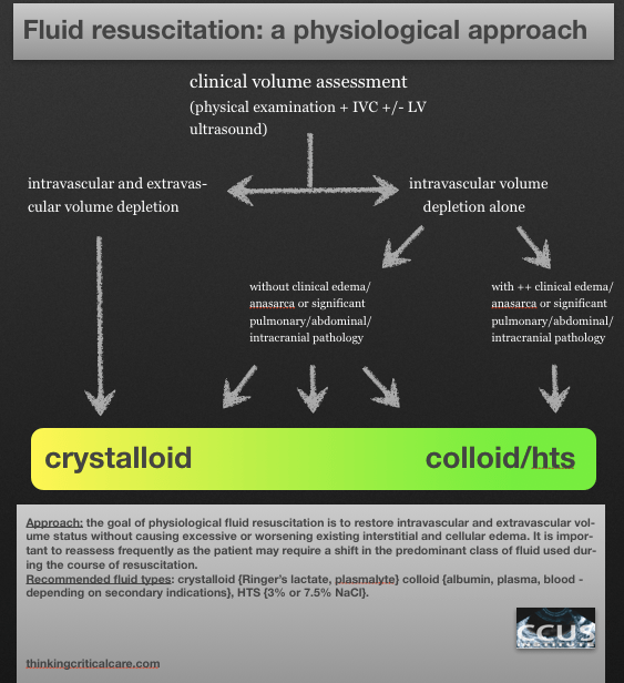
fluid resuscitation: a physiological approach – an N=1 podcast, #FOAMed, #FOAMcc

This is my approach to fluid resuscitation – sorry for the lack of precision which, to me, is actually key.  It would be against the N=1 principle to give out a recipe…but here’s a way to think about it:

Sorry the last bit cut off – my iphone can only email an 8 minute audio clip! Which I wasn’t aware of until today.  Anyway all that was lost at the end was “thanks for listening and I’d really like to hear comments and others’ practices!”

And here’s a disclaimer:  I don’t think this is the be-all and end-all. My resuscitation is a work in progress, both in terms of new fluids coming up, and in terms of identifying subgroups or individuals who would benefit from a different approach, so I’m definitely eager to hear from anyone who does things differently – but physiologically!

Please see Dr. John Myburgh’s excellent review on fluid resus in NEJM sep 26th issue!

Oh and here’s the diagram!

Physiological Fluids

thanks!

Philippe

Bedside Ultrasound Picture Quiz 5 – #FOAMed, #FOAMcc

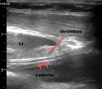
A 55 year old man admitted to the ICU for sepsis, recovering multi-organ failure with persistent culture-negative fever.

Longitudinal view of the left internal jugular vein.

What do you see?

BUPQ5-Q

scroll below for an answer!


BUPQ5-A


Thrombosed internal jugular catheter. This patient was anticoagulated. The fever disappeared within a few days and the thombosis decreased significantly.  It is difficult to be certain whether the fever was a cause but examining central lines is part of our fever workup.

Bedside Ultrasound Picture Quiz 2 #FOAMed, #FOAMcc

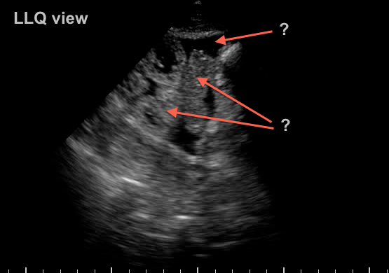
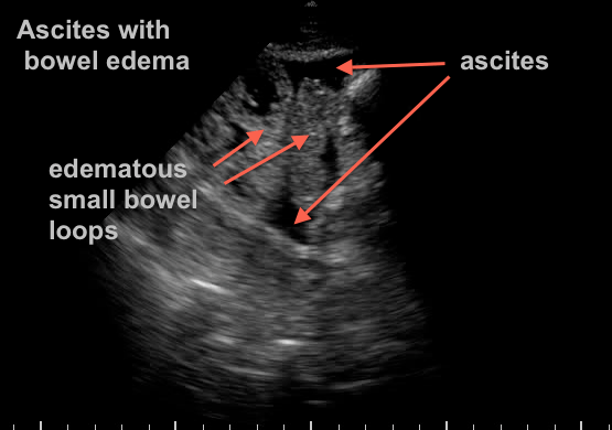
73 yr old woman recovering from septic shock with abdominal distension and difficulty tolerating enteral feeds…


what do you see?

BUPQ2


scroll below for the answer…..


BUPQ2 Answer


Extensive third spacing from resuscitation has resulted in bowel edema and ascites.  Another “benign” effect of massive crystalloid use… A bedside 22g US guided tap confirms benign transudate.

Case Studies in Bedside Ultrasound

I’ll soon start putting up interesting bedside ultrasound loops and images (as soon as I can get this software figured out…), in the meantime, if anyone is interested, we have a casebook  which can be found on the CCUS website (http://www.ccusinstitute.org or an e-version on iTunes (https://itunes.apple.com/us/app/50-case-studies-in-bedside/id599201706?mt=8).

thanks!

The Ideal Resuscitation Fluid – an N=1 Podcast #FOAMed, #FOAMcc

So here is the first in a series of mini-talks geared towards having us think about fluid resuscitation, which, for those in acute care of almost any type (ER, wards, ICU, anasthesia, surgery, etc…) is part of our daily routine. And that’s exactly what it shouldn’t be, routine. It should be carefully thought out and adapted to each individual clinical situation we’re facing.

So I’ve decided to approach this from a completely different angle, not looking at what we do, what’s available, and see what has been stacked up against what, etc, etc… Instead, I’ve decided to start the discussion from a completely theoretical standpoint and talking about something that doesn’t exist:  The Ideal Resuscitation Fluid.

Please, let me know your thoughts!

Philippe

http://www.ccusinstitute.org

(sorry the last 40 seconds were cut – now the “full” 5 minutes are up!  apologies, I am techno-challenged!)

How much fluid should I give? #FOAMed, #FOAMcc

I’m putting this question out there so that everyone can reach into their bag of tricks and tell me “this is how I know how much to give.”  Because I really do want to know.

To the best of my knowledge, that answer does not exist.

Of course, someone can say “we have to give 2 liters of crystalloids!” loudly and clearly, raising a fistful of papers and guidelines where it is printed, black on white.

Now lets think about exactly what that question is really asking, which is: how much fluid can I safely give my patient to improve the hemodynamic status without a significant cost in terms of complications, both short and long-term?

What are the implications of this statement?

a. My patient must be volume responsive.

b. I must not cross the threshold of excessive fluid administration. 

c. I must KNOW that fluid loading is the most appropriate therapy for my patient.

Now when you start looking at those questions and really wondering whether you have a definite answer for all of them, if becomes quite difficult.  It is much easier to say “well, such-and-such study says people do better if I give them x amount.”  This is probably true, that most people do better with a certain amount of fluid. But not everyone, because there are a lot of variables within those three questions.

Lets go through these questions:

My patient must be volume responsive – this is worth an entire podcast/lecture/article and there are a number of technologies and techniques out there which have both physiological and practical merit. I’ll get into that soon, but the short answer is that a passive leg raise (PLR) coupled with good cardiac output monitoring would be the “gold medal,” bedside ultrasound of the IVC’s global dynamics would be the “silver medal” and most of the rest would be “bronze medal” material at best. I won’t even mention the traditional physical exam or history or labs, not only do they not make it onto the podium, they didn’t make it into the stadium. I have to thank Scott Weingart (www.emcrit.org) for the sports analogy which I think is really excellent, since making the podium is actually pretty good and the difference between the three medalists may not always be that great.

I must not cross the threshold of excessive fluid administration – this one is dicey, because that’s where you have to factor in the patient’s oncotic pressure, hydrostatic pressure, both of which can be somewhat estimated, but mostly the degree of leak or “capillaritis”, both systemic and local, and that, at initial presentation, is impossible. The best you can do is a very broad classification from not too leaky to really, really leaky. And that’s not science. Now my good and highly esteemed friend Daniel Lichtenstein (the true founder of bedside ultrasound) published the FALLS Protocol, which basically says you can fill someone up until the appearance of B lines (during resuscitation) by lung ultrasound is certainly right that you should stop at this point, but I would contend you probably need to stop a bit before the edema is actually happening, especially since, in all likelihood and especially with crystalloids, the extravasation will continue for a while. The SOAP and VASST studies clearly showed an association between positive fluid balance and mortality, all other factors adjusted. Hence, this question is almost impossible to answer with knowledge. An educated guess is the best we can hope for.

I must KNOW that fluid loading is the most appropriate therapy for my patient – now this is almost philosophical and anxiety-arousing, because it forces us to challenge the fundamentals of our therapy. In certain cases, it is quite clear. If someone has been hemorrhaging or overdiuresed into shock, it is fair to say that you know they need fluids. However, if we are dealing with a vasodilatory circulatory failure, why do we feel compelled to treat it with a therapy best reserved for hypovolemic circulatory failure…  Hmmm. Food for thought. Perhaps we can start by looking back and seeing what the basis is for our almost unshakeable beliefs that fluids are benign and that vasoactive medications are evil… But that is for the near future.

Please, let me know what you think. I hope someone disagrees strongly.


Philippe Rola

http://www.ccusinstitute.org