Bedside Ultrasound Picture Quiz #6 – #FOAMed, #FOAMcc

So your intubated patient has developed abdominal distension and residuals are 250. You scan the LUQ:


Screen Shot 2013-10-28 at 11.32.45 PM


what do you see?  what should you do?

scroll below…


dilated stomach2


You have gastric distension with at least a liter of fluid in there.   Put in an OG, hold feeds, assess the rest of the abdomen to r/o bowel obstruction, consider motility agents and look for a reason behind the ileus or obstruction.


Philippe

CCUS 2013 Lectures – #FOAMed, #FOAMcc

This past may we had an amazing two day conference, the theme of which was challenging dogmatic practice and myths in acute care medicine.  Many of the lectures are now available to watch on our website at http://www.ccusinstitute.org/e-Store.asp?method=evideos#, you need to be a member to access – which is free, just register.


Lectures on bedside ultrasound, shock, ECMO in the ED, physiology and a lot of really, really good stuff.


We will be adding more in the next weeks!


Thanks!


PR

Bedside Ultrasound Picture Quiz 5 – #FOAMed, #FOAMcc

A 55 year old man admitted to the ICU for sepsis, recovering multi-organ failure with persistent culture-negative fever.

Longitudinal view of the left internal jugular vein.

What do you see?

BUPQ5-Q

scroll below for an answer!


BUPQ5-A


Thrombosed internal jugular catheter. This patient was anticoagulated. The fever disappeared within a few days and the thombosis decreased significantly.  It is difficult to be certain whether the fever was a cause but examining central lines is part of our fever workup.

Bedside Ultrasound Picture Quiz 2 #FOAMed, #FOAMcc

73 yr old woman recovering from septic shock with abdominal distension and difficulty tolerating enteral feeds…


what do you see?

BUPQ2


scroll below for the answer…..


BUPQ2 Answer


Extensive third spacing from resuscitation has resulted in bowel edema and ascites.  Another “benign” effect of massive crystalloid use… A bedside 22g US guided tap confirms benign transudate.

Bedside Ultrasound Picture Quiz 1 #FOAMed, #FOAMcc

A 70 year old man with cough and fever…what do you see?

Right costal coronal view


scroll down for an answer!


Rt CCV labelled


The ultrasound diagnosis is of a consolidation with a small pleural effusion.  This can be referred to a “hepatization” as the appearance becomes quite similar to an unaerated organ. A sonographic air bronchogram is seen as well. Clinical correlation with the history and bloodwork strongly suggests a pneumonia.

The Ideal Resuscitation Fluid – an N=1 Podcast #FOAMed, #FOAMcc

So here is the first in a series of mini-talks geared towards having us think about fluid resuscitation, which, for those in acute care of almost any type (ER, wards, ICU, anasthesia, surgery, etc…) is part of our daily routine. And that’s exactly what it shouldn’t be, routine. It should be carefully thought out and adapted to each individual clinical situation we’re facing.

So I’ve decided to approach this from a completely different angle, not looking at what we do, what’s available, and see what has been stacked up against what, etc, etc… Instead, I’ve decided to start the discussion from a completely theoretical standpoint and talking about something that doesn’t exist:  The Ideal Resuscitation Fluid.

Please, let me know your thoughts!

Philippe

http://www.ccusinstitute.org

(sorry the last 40 seconds were cut – now the “full” 5 minutes are up!  apologies, I am techno-challenged!)

How much fluid should I give? #FOAMed, #FOAMcc

I’m putting this question out there so that everyone can reach into their bag of tricks and tell me “this is how I know how much to give.”  Because I really do want to know.

To the best of my knowledge, that answer does not exist.

Of course, someone can say “we have to give 2 liters of crystalloids!” loudly and clearly, raising a fistful of papers and guidelines where it is printed, black on white.

Now lets think about exactly what that question is really asking, which is: how much fluid can I safely give my patient to improve the hemodynamic status without a significant cost in terms of complications, both short and long-term?

What are the implications of this statement?

a. My patient must be volume responsive.

b. I must not cross the threshold of excessive fluid administration. 

c. I must KNOW that fluid loading is the most appropriate therapy for my patient.

Now when you start looking at those questions and really wondering whether you have a definite answer for all of them, if becomes quite difficult.  It is much easier to say “well, such-and-such study says people do better if I give them x amount.”  This is probably true, that most people do better with a certain amount of fluid. But not everyone, because there are a lot of variables within those three questions.

Lets go through these questions:

My patient must be volume responsive – this is worth an entire podcast/lecture/article and there are a number of technologies and techniques out there which have both physiological and practical merit. I’ll get into that soon, but the short answer is that a passive leg raise (PLR) coupled with good cardiac output monitoring would be the “gold medal,” bedside ultrasound of the IVC’s global dynamics would be the “silver medal” and most of the rest would be “bronze medal” material at best. I won’t even mention the traditional physical exam or history or labs, not only do they not make it onto the podium, they didn’t make it into the stadium. I have to thank Scott Weingart (www.emcrit.org) for the sports analogy which I think is really excellent, since making the podium is actually pretty good and the difference between the three medalists may not always be that great.

I must not cross the threshold of excessive fluid administration – this one is dicey, because that’s where you have to factor in the patient’s oncotic pressure, hydrostatic pressure, both of which can be somewhat estimated, but mostly the degree of leak or “capillaritis”, both systemic and local, and that, at initial presentation, is impossible. The best you can do is a very broad classification from not too leaky to really, really leaky. And that’s not science. Now my good and highly esteemed friend Daniel Lichtenstein (the true founder of bedside ultrasound) published the FALLS Protocol, which basically says you can fill someone up until the appearance of B lines (during resuscitation) by lung ultrasound is certainly right that you should stop at this point, but I would contend you probably need to stop a bit before the edema is actually happening, especially since, in all likelihood and especially with crystalloids, the extravasation will continue for a while. The SOAP and VASST studies clearly showed an association between positive fluid balance and mortality, all other factors adjusted. Hence, this question is almost impossible to answer with knowledge. An educated guess is the best we can hope for.

I must KNOW that fluid loading is the most appropriate therapy for my patient – now this is almost philosophical and anxiety-arousing, because it forces us to challenge the fundamentals of our therapy. In certain cases, it is quite clear. If someone has been hemorrhaging or overdiuresed into shock, it is fair to say that you know they need fluids. However, if we are dealing with a vasodilatory circulatory failure, why do we feel compelled to treat it with a therapy best reserved for hypovolemic circulatory failure…  Hmmm. Food for thought. Perhaps we can start by looking back and seeing what the basis is for our almost unshakeable beliefs that fluids are benign and that vasoactive medications are evil… But that is for the near future.

Please, let me know what you think. I hope someone disagrees strongly.


Philippe Rola

http://www.ccusinstitute.org

Why do we bother checking CVP? #FOAMed, #FOAMcc

I was recently scanning the literature in preparation for our symposium, and came across what should have been a 2003 instead of a 2013 publication in the March issue of the CCM Journal, entitled “Point-of-Care Ultrasound to estimate Central Venous Pressure:  A Comparison of Three Techniques.”

I have to admit this is a pet peeve of mine, from the standpoint of a clinical physiologist, which is, as far as I’m concerned, what any physician looking after critically ill patients should be, at least some of the time.

So our real question is: is my patient fluid-responsive?  And perhaps a corollary question would be: is he fluid tolerant?

As a longtime bedside sonographer, physiology, experience and slowly growing evidence all support my using IVC sonography as a tool to assess volume responsiveness.  It isn’t perfect, and personally, I find the common M-mode, two-point measurement to be inadequate compared to a global assessment of the IVC, but it certainly is far closer to “the truth” we seek than CVP.

This then begs the question: why on earth would we be seeking to correlate one type of data to another which is clearly more removed from “the truth” we seek?

The use of CVP is largely cultural and deeply ingrained. There are some limited ways and pathologies in which it can be useful, but not as a measure of preload.  My friend Paul Marik published a piece that was both enlightening and entertaining in Chest a couple of years ago which I would have thought would have been the final nail in the coffin for the use of CVP as a preload tool, but it endures…even in the latest surviving sepsis guidelines

A testament to religion over science.

Philippe Rola

http://www.ccusinstitute.org

note that this was first posted in my buddy Matt’s awesome website pulmccmcentral (http://pulmccm.org/2013/critical-care-review/why-do-we-bother-to-check-cvp/) please check it out!

Surviving Sepsis Guidelines: useful, but patients deserve individualized care! #FOAMed, #FOAMcc

First of all, I would like to commend those involved in the Surviving Sepsis Campaign’s Guidelines. It is a tremendous endeavour that, without a doubt, has heightened awareness and their growing implementation has and will save many lives.

I would, however, also like to point out that guidelines are exactly what the term implies, and not necessarily a gold standard to  aspire to and adhere to in religious fashion.  The reason this is so is the inherent variability in human physiology and pathology.  If, out of 100 patients a treatment would help 10 but harm 1, the numbers and studies would clearly support its broad use. We’d win more than we’d lose. However, as physicians, we treat the one patient in front of us, not the hundred, so I find it difficult to believe that such blind application of a recipe would be the most Hippocratic practice to apply.

We know that our patients are widely different, and around the corner is point of care immunology that will tell us, in all likelihood, that even patients we think are similar on the surface will have widely different immune profiles and will respond to treatment differently.

There are a few recommendations which, to me, make little physiological sense, particularly in certain circumstances:

I don’t think it necessary to belabour the point about CVP. As a static measure, CVP has clearly been disproven to have any relevance in predicting volume responsiveness. Its persistence is a testament to cultural faith rather than science.

I would find it unethical to blindly bolus large amount of crystalloids (which we all know end up 70-80% extravascular) in a patient when it takes about 10 seconds to rule out a pre-existing, septic cardiomyopathy or a volume-intolerant state. Even more so when dealing with pathologies where third-spacing is a concern (pulmonary and intra-abdominal pathologies) since those leaky capillaries is where most of that fluid resuscitation will end up. Yes, I am implying that a worsening chest x-ray is not always and only due to worsening disease… Physiologically, perhaps non synthetic colloids or hypertonics may be a better option…

I would find it equally unethical to blindly put a patient on dobutamine who may have hyperdynamic ventricles and possibly still a volume-responsive state. Again, determining this takes seconds.

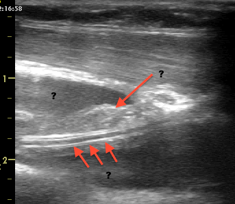
Yes, clearly I admit to a bedside ultrasound bias. It allows us to look inside our patients. Isn’t that what we’re always trying to do?

I can already hear voices and keyboards claiming the “lack of evidence,” and they would partly be right. That evidence is slowly but surely growing.

Unfortunately, point of care ultrasound has come of age in the era of evidence based medicine, and, as such, is required to “pass” that scrutiny whereas most of what is currently being done was “grandfathered in” and given a bye. I would be interested in seeing the compelling evidence for the use of a stethoscope.

The evolution of evidence based medicine is an interesting scientific, commercial and social development. From the positive study publishing bias to the general lack of epidemiological knowledge of our community, and without mentioning the darker side of research and publishing, it is unfortunate that almost every statement by a physician, to be taken seriously, must be backed by a hand raising a publication. And how many of those do we see torn down a month, a year or five later, thoroughly disproven? The pendulum of evidence-based medicine has perhaps swung too far…

Note that I’m not trying to discredit the countless number of truly well-designed and well-executed studies that contribute immensely to medicine – which would otherwise be reduced to little more than expert opinion – just that careful analysis of both the evidence and the case at hand is primordial.

I think that as physicians, it is our duty to look very closely at the individual patient, the care of whom we are privileged to have as a responsibility, and individualize our treatment plan to his specific problem given his specific physiology, and not blindly implement a recipe, even if it would happen to be the right one 9 out of 10 times.

Philippe Rola

http://www.ccusinstitute.org

please note that this was first posted on september 5th, 2013 on my buddy Matt’s website, pulmccm.org (http://pulmccm.org/2013/uncategorized/surviving-sepsis-guidelines-useful-patients-deserve-individualized-care/)

Go visit, its great, and you can see some fun follow up comments, too!

Glenn says:

it’s a sensitive decease in a sensitive time. Time is of the essence in treating septic shock and severe sepsis. If you wait for primary MD to individualized the care for these patients,it’s probably too late.EGDT save lives.

Thanks for commenting Glenn, and I can’t agree more. EGDT does save lives when compared to the usual care of 2001 (see my latest post on the ProCESS trial as that may no longer be the case in some institutions), but the mistake is strict adherence when you do have the capabilities to detect conditions where a protocol “violation” would be beneficial to the individual patient in front of you. Again, I’m not knocking EGDT, it was a great step, but in a set of stairs we have to keep climbing.


Philippe