The Glycocalyx: an overview for the clinician. #FOAMed, #FOAMcc

Ok, so I’d had a couple of glimpses at articles in the past few years which referred to the glycocalyx, but, in truth, I tend to read most of the “bench” studies a little, well…quickly.  So basically, when I listened to Paul’s (Marik) recent lecture at Scott’s (Weingart, emcrit.org) New York Sepsis Collaborative, I started to dig a little…and whoa! And then of course, then now-famous expose by John (Doe?) on EmCrit continued to convince me that this is definitely something I need to pay attention to! Its not like there hasn’t already been a high level of scrutiny of the glycocalyx in the field of sepsis.  Google it. Its like a whole new world. It just hasn’t yet translated into an effective therapy, but nor has it seemed to spread into general awareness, and it seems like it’s high time it does, since it is the interface between the blood and the body – a “blood-body barrier” of sorts.

So here we go, a crash course on the glycocalyx for the clinician in the trenches…

The existence of an acellular layer lining the endothelium was described by Luft some 40 years ago (1), and in the last decade has come under scrutiny for its role in various pathophysiological states, which seems to be quite exhaustive. This has not yet translated into diagnostic or therapeutic interventions, but it seems its properties, or at least those we are currently aware of, should be kept in mind when we are faced with therapeutic choices given that some of these may have an effect on the glycocalyx.

Its existence was deduced due to the lower capillary hematocrit – meaning that the hematocrit in the capillaries, adjusted for the luminal volume, is lower than that of the large or medium vessels, implying an area where there are no red cells. This was confirmed by electron microscopy and found to be a gel-like epithelial lining which acts as an interface between the blood and the endothelial cells, of a thickness of about 0.5 um at the level of tha capillaries, and thicker in the larger vessels.

So what is it made of? Its is essentially a meshwork of glycoproteins and proteoglycans, anchored to the epithelial cells, in which many soluble molecules are enmeshed. It is important to note that there is a dynamic equilibrium between this layer and the adjacent flowing blood, which will affect the thickness and composition of the glycocalyx. The layer seems to be vulnerable to both enzymatic degradation as well as to shear forces, in variable degrees. Enzymatic removal of components seems to radically alter properties, pointing to a strong synergistic effect of the various components. It is a constantly shedding and regenerating structure.

glycocalyx em pic

EM view of the glycocalyx (reproduced with authors’ permission from Reitsma et al.)

glycocalix pic2

(reproduced with authors’ permission from Reitsma et al.)

Major components:

– Proteoglycans (protein core with chains of glycosaminoglycans) are the “backbone” of the glycocalyx, and consist of syndecans, glipicans, mimecan, perlecans and biglycans.

– Glycosaminoglycans (linear disaccharide polymers of a uronic acid and a hexosamine) are predominantly heparan sulfate (50-90%), then dermatan sulfate, chondroitin sulfate, keratan sulfate and hyaluronan (or hyaluronic acid).

– Glycoproteins are also part of the “backbone” structure and the main types are the endothelial cell adhesion molecules ( -cams, which are selectins, integrins and immunoglobulins) and components of the fibrin/coagulation system.  E- and P-selectins as well as others are involved in leukocyte-endothelial interaction and diapedesis, an important aspect of local inflammation.

– Soluble components are also embedded in the glycocalyx such as proteins and soluble proteoglycans and are important in preserving the charge of the layer and play critical roles in functionality.

Function of the glycocalyx (as far as we know…)

a. gatekeeper

It is a key determinant of vascular permeability. Partial enzymatic removal without damage to the endothelial cells themselves result in a radical change in permeability in aminal models. Charge, size and steric hindrance affect permeability. The glycocalyx has a highly net negative charge towards the bloodstream – neutralizing this induces an increase in cellular albumin uptake.

Weinbaum introduced a new model integrating the glycocalyx in the classical (but now outdated and disproven) Starling model of microvascular fluid exchange. The revised Starling principle stresses the importance of an intact glycocalyx.

Its role with cellular elements is interesting, as it contains key elements for interaction (-CAMs) but at the same time physically prevents direct interaction between cells (WBC, RBC, plt) and the endothelium. This clearly points to a pivotal role in controlling the interaction. Damage by various methods consistently shows increased neutrophil-endothelial interaction (often termed “leukocyte rolling”). It isn’t much of a stretch to see how the glycocalyx will thus be involved in the control of local inflammation.

b. mechanotransduction

The glycocalyx provides mechanical protection from shear stress to the endothelium.  Increased shear leads to upregulation of synthesis, and correspondingly, thicker glycocalyx is found in high shear areas.

c. microenvironment

Receptor binding, local growth and repair, and vasculoprotection. For instance, ATIII is bound to the glycocalyx (inhibits procoagulants), as well as superoxide dismutase, key in reducing oxidative stress and maintaining MO availability.

Clinical implications…

Now this is the real question.  I think that the first step is acknowledging the presence and importance of the glycocalyx, and trying to discern which of our interventions may have an impact.  It is quite clear that enzymes, cytokines and ischemia/reperfusion all damage the glycocalyx and result in increased cellular interaction and permeability. In a way this can explain the entire “SIRS” spectrum with diffuse damage resulting from an insult that may or may not be infectious in origin. Obviously, we know to avoid anything that might cause the above.

I think we can divide our interventions into two types:

a. those that inherently disrupt the glycocalyx –

b. those that secondarily disrupt it via another mediator – eg over-resuscitation and ANP/BNP (John’s “evil twins”) elevation causing breakdown.

Here are some interesting facts, in no particular order of importance:

a. in acute hyperglycemia and in type I diabetes, there is significant loss of glycocalyx volume.

Nieuwdorp M, van Haeften TW, Gouverneur MC, Mooij HL, van Lieshout MH, Levi M, Meijers JC, Holleman F, Hoekstra JB, Vink H, Kastelein JJ, Stroes ES (2006) Loss of endothelial glycocalyx during acute hyperglycemia coincides with endothelial dysfunction and coagulation activation in vivo. Diabetes 55:480–486.

Nieuwdorp M, Mooij HL, Kroon J, Atasever B, Spaan JA, Ince C, Holleman F, Diamant M, Heine RJ, Hoekstra JB, Kastelein JJ, Stroes ES, Vink H (2006) Endothelial glycocalyx damage coincides with microalbuminuria in type 1 diabetes. Diabetes 55:1127–1132

b. high fat/high cholesterol diet and high LDL levels disrupt the glycocalyx. The co-infusion of superoxide dysmutase and catalase with ox-LDL abolishes the glycocalyx shedding found with ox-LDL infusion only. (not exactly a big surprise, but certainly brings up a therapeutic possibility in acute coronary events, especially NSTEMIs…)

Vink H, Constantinescu AA, Spaan JA (2000) Oxidized lipoproteins degrade the endothelial surface layer: implications for platelet–endothelial cell adhesion. Circulation 101:1500–1502

van den Berg BM, Spaan JA, Rolf TM, Vink H (2006) Atherogenic region and diet diminish glycocalyx dimension and increase intima-to-media ratios at murine carotid artery bifurcation. Am J Physiol Heart Circ Physiol 290:H915–H920

c. hydrocortisone and antithrombin prevent TNF-a induced shedding of the glycocalyx. (we never really knew how steroids help in sepsis; ATIII trials failed, but were they designed with glycocalyx-sparing in mind…?)

Daniel Chappell MD, Klaus Hofmann-Kiefer, Matthias Jacob, Markus Rehm, Josef Briegel, Ulrich Welsch, Peter Conzen, Bernhard F. Becker TNF-α induced shedding of the endothelial glycocalyx is prevented by hydrocortisone and antithrombin Basic Research in Cardiology, January 2009, Volume 104, Issue 1, pp 78-89

 d. endotoxemia damages the pulmonary vascular endothelium and results in ALI (now the neutrophils can attach and activate inflammation…)

Eric P Schmidt, Yimu Yang, William J Janssen, Aneta Gandjeva, Mario J Perez, Lea Barthel, Rachel L Zemans, Joel C Bowman, Dan E Koyanagi, Zulma X Yunt, Lynelle P Smith, Sara S Cheng, Katherine H Overdier, Kathy R Thompson, Mark W Geraci, Ivor S Douglas, David B Pearse & Rubin M Tuder The pulmonary endothelial glycocalyx regulates neutrophil adhesion and lung injury during experimental sepsis, Nature Medicine 18,1217–1223 (2012)

e. sepsis and major abdominal surgery damage the glycocalyx (a good example of common pathway pathophysiology…)

Jochen Steppan, M.D., Stefan Hofer, M.D., Benjamin Funke, M.D., Thorsten Brenner, M.D., Michael Henrich, M.D., Ph.D., Eike Martin, M.D.,Jürgen Weitz, M.D., Ursula Hofmann, M.D., Markus A. Weigand, M.D.Sepsis and Major Abdominal Surgery Lead to Flaking of the Endothelial Glycocalix Journal of Surgical Research Volume 165, Issue 1 ,Pages 136-141, January 2011

f. molecules (eg Slit2N,  good old aPC) that enhance or preserve barrier function have shown some success in animal models. (the key might be to using a bunch of them at once, not one of them vs placebo)

N. R. London, W. Zhu, F. A. Bozza, M. C. Smith, D. M. Greif, L. K. Sorensen, L. Chen, Y. Kaminoh, A. C. Chan, S. F. Passi, C. W. Day, D. L. Barnard, G. A. Zimmerman, M. A. Krasnow, D. Y. Li, Targeting Robo4-dependent Slit signaling to survive the cytokine storm in sepsis and infl uenza. Sci. Transl. Med. 2, 23ra19 (2010).

…and this is just the tip of the iceberg. Seriously, google it…

Conclusion:

For now, it is difficult to make any hard recommendations, but it has certainly made me pause to regroup and re-strategize. I think the critical thing is to reframe our thinking and redesign our approach to be a glycocalyx-sparing therapy.

In sepsis therapy, so many molecules have failed, but little emphasis so far has been on targeting the glycocalyx, and in all likelihood, the key is not in finding the “magic bullet” but rather using multiple interventions to bolster it. One could think of this as the sepsis “chain-of-survival”, and now that we can see the complexity of the glycocalyx, it is easy to understand how no one therapy, even if it did do its job, would succeed in preventing the degradation of the other links in the chain, and fail.  In all likelihood, a successful strategy will probably involve the following:

1. fancy molecules to prevent glycocalyx damage.

2. fluid choices which are glycocalyx-friendly.

3. fluid in just the right amount (not by macro/volume-responsiveness but by micro/glycocalyx management).

4. rapid diagnosis/abx/source control, etc, all the good stuff we know about.

5. preventing hyperglycemia.

A trial like that would be a monumental undertaking. I can only hope someone does it.

My next step, as a guy in the bedside trenches and not at the bench, after gaining a modicum of understanding on the topic, will be to delve deeper into the effects of currently available fluids on the glycocalyx.  Look for a post on that in the next weeks.  As a starter, everyone should review Woodcock & Woodcock’s excellent clinical review.

And if anyone has any amazing information to share, please do!!! That’s what #FOAMed is for!!!

Suggested viewing/reading:

Woodcock and Woodcock, BJA 2012,  http://bja.oxfordjournals.org/content/early/2012/01/29/bja.aer515.full.pdf+html

Broken Barriers: A New Take on Sepsis Pathogenesis Neil M. Goldenberg,1* Benjamin E. Steinberg,1* Arthur S. Slutsky,2,3,4 Warren L. Lee2,4 Science Translational Medicine, 22 June 2011 Vol 3 Issue 88 88ps25

Click to access Science_Translational_Medicine_2011_Pathogenesis_of_Sepsis.pdf

a great article on understanding the glycocalyx in sepsis by a U of T gang!

emcrit:

http://emcrit.org/blogpost/best-fluids-comment-ever/

http://emcrit.org/podcasts/fluids-severe-sepsis/

http://emcrit.org/podcasts/paul-marik-fluids-sepsis/

all must-listen/watch material!

other refs.

Luft JH (1966) Fine structures of capillary and endocapillary layer as revealed by ruthenium red. Fed Proc 25:1773-1783

Reitsma et al, The endothelial glycocalyx: composition, functions, and visualization, Eur J Physiol (2007) 454:345–359

Zhang X, Adamson RH, Curry FR, Weinbaum S (2006) A 1-D model to explore the effects of tissue loading and tissue concentration gradients in the revised Starling principle. Am J Physiol Heart Circ Physiol 291:H2950–H2964
Is it still cool to cool in cardiac arrest? The new TTM Hypothermia trial! #FOAMed, #FOAMcc

So the other dayI read the TTM trial (http://www.nejm.org/doi/full/10.1056/NEJMoa1310519?query=featured_home#t=article) with great interest, as cardiac arrest and the post-resuscitation phase have always been among my pet topics.

First of all this is a big trial.  Bigger that the previous ones that established hypothermia as a standard of care. Does it obviate those previous results?  Absolutely not. Those trials were not 32/33 vs 36 but 32/33 vs “whatever happens.”

Hypothermia makes a lot of sense physiologically, but of course that doesn’t mean that it might not have some harmful side effects that have not yet been clearly delineated (besides the current known hemodynamic ones and relatively benign electrolyte and renal alterations).

However, it is pretty clear that, compared to 33 degrees, 36 does just as well, which leans towards saying that all we have to do is avoid fever, or stay in a very mild hypothermia.

Avoiding secondary injury in brain pathology is key (no desat, no hypotension, and no fever), and in anoxic encephalopathy, it is no different.  The key thing is that in this trial, the temperature was controlled – ie it would not be acceptable to do no cooling, and just chase the fever (which is very common) with acetaminophen, which would invariably result in significant time spent above 36 (oops, tylenol didn’t really work, ok lets put the blanket, etc …this is gonna be hours).

So is this the end of aggressive cooling?  Not necessarily.

For anyone interested in the topic, I suggest reviewing Peter Safar‘s data on dogs and cold aortic flushes – it is absolutely unbelievable to see dogs who  had an arrest, got the cold aortic flush (brain temp below 10 degrees), are left stone cold dead for 45 minutes, then resuscitated and are then able to go around a few days later and do doggie things like run and bark and eat…  So I don’t think that cooler isn’t necessarily better, but that we haven’t yet delineated what are the pros and cons of each temperature range or how to get there practically and safely.

So what should we do?  Well, it would seem reasonable to do either at this point, and accepting a temp between 32-36 (I have usually preferred 33-34 as they rarely drift down into the 20’s as I’ve seen the 32’s do) as being adequate. This may make hemodynamics a bit easier to manage in certain cases.
Also check out Scott’s take at:
 http://emcrit.org/podcasts/emcrit-wee-targeted-temperature-trial-changes-everything/#comment-58635
And the RAGE Podcast addresses this topic at about 25 minutes:
…and of course, keep abreast of further data and subgrouping that may become available on this, and further trials. But for now, its definitely still cool to cool, maybe just a little less…

A Paradigm shift: re-thinking sepsis, and maybe shock in general… #FOAMed, #FOAMcc

Thomas Kuhn, physicist and philosopher, in his groundbreaking and science changing text, The Structure of Scientific Revolutions, states that:

“Successive transition from one paradigm to another via revolution is the usual developmental pattern of a mature science.”

In other words, a science has growing pains and is bound to have a fair bit of debate and controversy, until a new paradigm becomes dominant.  I think that there is a current – in part prompted by the power of socio-professional media which has allowed minds to connect and knowledge to spread – that will see many of the things that are now “Standard of Care” out the door.

So first of all, the following are must-listens, the first a lecture by Paul Marik, whom I have had the chance to collaborate with in the last years and respect greatly, on knowledge, experience, and even more on his refusal to take anything for granted and being in a seemingly-constant quest for the improvement of medicine.

The second link is Scott Weingart’s take on it, which I think is equally awesome.

I think Paul is pushing the envelope in an essential way, and Scott does a fantastic job of seeing or putting it in perspective. Enjoy:

http://emcrit.org/podcasts/paul-marik-fluids-sepsis/

EMCrit 112 – A Response to the Marik Sepsis Fluids Lecture

My (very) humble opinion on this is a rather simple, almost philosophical one:  why are we seemingly obsessed with treating a predominantly vasodilatory pathology with large amounts of volume?  I’ve said this in previous posts and podcasts, but this, in my opinion, is largely cultural and dogmatic. “Levophed – Leave’em dead” is something I heard as a student and resident, and came to take for granted that I should give lots of fluid in hopes of avoiding pressors… But there’s no evidence at all to support this.  The common behavior of waiting until someone has clearly failed volume resuscitation before starting pressors befuddles me (think how long it takes to get two liters of fluid in most ERs…).  If I was in that bed, I’d much rather spend an hour a bit “hypertensive” (eg with a MAP above 70) than a bit hypotensive while awaiting final confirmation that I do, in fact, need pressors.

I strongly suspect that it’s just a matter of improving vascular tone, giving some volume (which may be that 3 liter mark), and ensuring that the microcirculation/glycocalyx is as undisturbed as possible. Now when I say it may be the 3 liters, I firmly believe this will not apply to everyone, and that it will be 1 liter in some, and 4 in others, and that a recipe approach will be better than nothing, but likely harm some.

I think that blind (eg no echo assessment) of shock is absurd, and for anyone to propose an algorithm that does not include point-of-care ultrasound is only acceptable if they are in the process of acquiring the skill with the intention of modifying their approach in the very near future.

The whole microcirculation/glycocalyx is absolutely fascinating stuff, and undoubtedly will come under scrutiny in the next few years, and it is definitely something I will focus on in upcoming posts & podcasts. Our resuscitation has been macro-focused, and certainly it is time to take a look at the little guys, who might turn out to have most of the answers. For instance, there is some remarkable data on HDAC inhibitors (common valproic acid) and their salutatory effects in a number of acute conditions such as hemorrhagic shock (Dr. Alam) which have nothing to do with macro-resuscitation, and everything to do with cell signaling and apoptosis. Hmmm…

please share your thoughts!

thanks

Philippe

Steroids for cardiac arrest…really? My take on the VSE study – #FOAMed, #FOAMcc

So I’ve been asked a few times for my opinion about the VSE study in the last couple of months, so here we go.

JAMA. 2013;310(3):270-279. doi:10.1001/jama.2013.7832.

First of all, lets look at it from a theoretical perspective.  How would steroids contribute to ROSC (return of spontaneous circulation)?  Hard to believe they possibly could, given the ultra-short timeframe to ROSC – minutes mostly – and the much longer action of steroids.  However, it is quite possible – and in view of this study perhaps likely – that there is an effect on shock and RONF (return of neurological function).

Why?  Post arrest shock results in MSOF due to a cascade of inflammation resulting from the hypoxic insult. Remember that we are not designed to survive these events. Being designed to fight off moderate trauma and infection (eg being bitten by an animal or clubbed by another caveman) our physiological reaction often overshoots the mark resulting in more damage than good, as it does in sepsis (variably depending on our different geno/phenotypes).  So whether liver, kidney or brain damage, some component is not only related to pure hypoxia but also to an inflammatory cascade that has a prolonged effect. This is the same thing we are targeting with cooling, on top of a simple metabolic supply/demand issue, so in terms of biological plausibility, it makes some sense.

In the post-ROSC phase, there is always the possibility of relative adrenal insufficiency – after all, the adrenals have taken a hit as all the other organs did – so again there is biological plausibility.

There’s quite a bit of debate out there as to whether or not to apply this.  I’m pragmatic, not a purist, and my beliefs lie in evidence, biological plausibility and the risk/benefit ratio.  In this case, I think the decision is actually quite simple.  The way I see it, the steroids are harmless and probably helpful, so I have been giving solumedrol in the last few months.

If anything, I’m more concerned about the harm I may be doing with epinephrine/vasopressin, especially in terms of RONF.  I do hope an epi (various doses) vs placebo study is done, because it is difficult to withhold, knowing that there is greater immediate effect on ROSC… Hard decision as the clinician at the bedside, and hopefully this will become clearer in the near future.

For those unclear about the whole epi debate, the physiological issue is that the relationship between pressure and perfusion is represented by an inverted U curve – at very high pressures (from vasoconstriction) perfusion is decreased (think of the extremities on high dose pressers with a decent BP).  So although we may help coronary perfusion pressure and thus ROSC, end-organ damage is greater…and nothing matters much without a brain.


So bottom line:  I’d go ahead with the steroids, and for now the V and E, but I wouldn’t be surprised to drop or decrease those soon.

More to come on resuscitation and its future (the present for some of us…) in posts and podcasts!

Hope this helps!

Philippe


CCUS Annual Symposium 2014 – The Ultrasound-Assisted Physical Exam! stay tuned!

This year, we’re putting together a really, really interesting event.  Bedside ultrasound being a hot topic and at the brink of revolutionizing clinical examination and practice, we figured that this year, we’d go back to basics to some degree with a general ultrasound approach, but also a step further in looking at it from an integration perspective, meaning how to approach clinical problems with ultrasound as an added tool.

Talks will be clinical problem-based – e.g. the patient with dyspnea, the patient with renal failure, etc, essentially showing participants how to integrate their growing ultrasound skills into routine use.  There will be a ton of faculty led workshops to review all the basic ultrasound skills (lung, cardiac, abdominal, vascular) on live models and on advanced CAE simulators, both adult and pediatric.

Our faculty will be fantastic, including Andre Denault, Haney Mallemat (@criticalcarenow), JF and Max (@EGLS_JFandMax), Edgar Hockmann, Catherine Nix, Alberto Goffi, Massimiliano Meineri, Matt Hoffmann (www.pulmccm.org), Jeff Burzynski, Jason Fisher, Alyssa Abo and many more…

The two day core event will take place in Montreal, on may 10th and 11th, and, equally interesting will be a pre-congress set of courses on may 9th, including:

EGLS (echo-guided life support)

Focused TEE

Bedside ultrasound for nurses (vascular access, IVC volume status assessment)

Critical Care Procedures (drains, tracheostomy, central lines)


Registration is not yet open but will be in the next few weeks, so for anyone interested please visit http://www.ccusinstitute.org and join (it’s free!) and we’ll email you when its up and running.

Please forward/link this to all your forward-thinking colleagues~

thanks!


Philippe

CCUS 2013 Lectures – #FOAMed, #FOAMcc

This past may we had an amazing two day conference, the theme of which was challenging dogmatic practice and myths in acute care medicine.  Many of the lectures are now available to watch on our website at http://www.ccusinstitute.org/e-Store.asp?method=evideos#, you need to be a member to access – which is free, just register.


Lectures on bedside ultrasound, shock, ECMO in the ED, physiology and a lot of really, really good stuff.


We will be adding more in the next weeks!


Thanks!


PR

Bedside Ultrasound Clip Quiz 2 – #FOAMed, #FOAMcc

64 yr old woman POD#3 Rt heimcolectomy with fever and abdominal pain.

Right costal longitudinal scan. What do you see?


A complex echogenic sliver of ascites over the liver…now what do you do? Here’s what I did…


…and it showed fecaloid material. Turned out to be anostomotic leak.  Didn’t have to push much for surgery.


Philippe


Bedside ultrasound clip quiz 1 – #FOAMed, #FOAMcc

62 year old with weight loss, tachycardia and progressive dyspnea…what do you see?

scroll below!


This was a case of pre-tamponade/tamponade.  Here is a view a few minutes later, with the guidewire in. This was a case of malignant pericardial effusion.

fluid resuscitation: a physiological approach – an N=1 podcast, #FOAMed, #FOAMcc

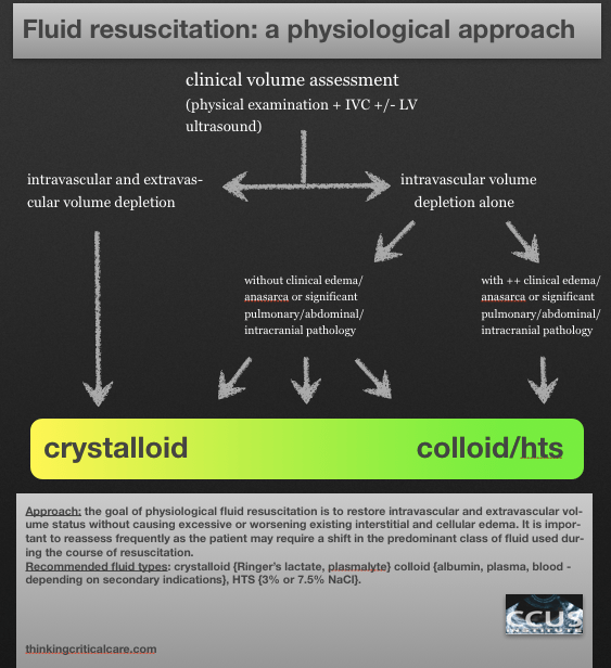
This is my approach to fluid resuscitation – sorry for the lack of precision which, to me, is actually key.  It would be against the N=1 principle to give out a recipe…but here’s a way to think about it:

Sorry the last bit cut off – my iphone can only email an 8 minute audio clip! Which I wasn’t aware of until today.  Anyway all that was lost at the end was “thanks for listening and I’d really like to hear comments and others’ practices!”

And here’s a disclaimer:  I don’t think this is the be-all and end-all. My resuscitation is a work in progress, both in terms of new fluids coming up, and in terms of identifying subgroups or individuals who would benefit from a different approach, so I’m definitely eager to hear from anyone who does things differently – but physiologically!

Please see Dr. John Myburgh’s excellent review on fluid resus in NEJM sep 26th issue!

Oh and here’s the diagram!

Physiological Fluids

thanks!

Philippe

Bedside Ultrasound Picture Quiz 2 #FOAMed, #FOAMcc

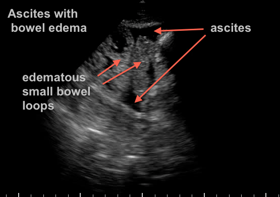
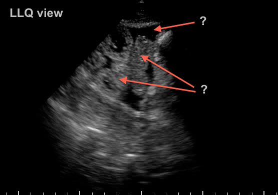
73 yr old woman recovering from septic shock with abdominal distension and difficulty tolerating enteral feeds…


what do you see?

BUPQ2


scroll below for the answer…..


BUPQ2 Answer


Extensive third spacing from resuscitation has resulted in bowel edema and ascites.  Another “benign” effect of massive crystalloid use… A bedside 22g US guided tap confirms benign transudate.