Bedside Ultrasound Clip Quiz: Abdominal pain and fever! #FOAMed, #FOAMcc, #FOAMus

Saw this poor fellow recently who presented to the ED with fever and abdominal pain. 73 years young. He came to my attention because of borderline BP (95 systolic) and a lactate of 4.5 mmol.

Here is a transverse scan at his lower right costal margin:


What do you think?

Turns out he had been having pain for about two weeks, and it had intensified about two days ago. His wife dragged him in.

What would you do?


This is septated fluid collection around the liver.  With the fever and history, sounds pretty suspicious for a septic source. After carefully scanning in all angles and watching for a while to make sure this wasn’t a strangely placed loop of bowel, a 22g needle aspiration showed cloudy bilious fluid and a trip to the OR a couple of hours later revealed a perforated duodenal ulcer.

He made it ok.


cheers


Philippe

L’Échographie au chevet pour l’Interniste: ASMIQ/CCUS 2014, #FOAMed, #FOAMcc, #FOAMus

Belle journée aujourd’hui à a deuxième collaboration éducative entre l’ASMIQ (Association des Spécialistes en Médecine Interne du Quebec) et le CCUS, lorsque une trentaine d’internistes ont approfondi leur habiletés échographiques.

Tel que promis, et dans l’esprit de #FOAMed, voici les présentations et videos:

Dr. Ian Ajmo nous fait une revue de l’évaluation de la volémie, en particulier l’échographie de la veine cave inférieure (VCI):

présentation PDF gestion-voleěmie – copie

vidéo https://vimeo.com/96308958,

Dr. Anne-Patricia Prévost révise l’échographie pulmonaire et cardiaque ciblée:

presentations PDF ASMIQ 2014 coeur ASMIQ 2014 poumons

vidéos:

https://vimeo.com/96315383,         https://vimeo.com/96315382,


Merci aussi à Dr. Simon Benoit et Dr. Nicolas Buissières qui ont fait un travail excellent dans les ateliers pratiques!


Philippe


Bedside Ultrasound for Internists / L’Échographie Clinique pour l’Interniste – ASMIQ/CCUS, May 23rd, 2014.

Although bedside ultrasound truly started in the emergency room and is slowly spreading to critical care, its usefulness extends to all aspects of medicine, particularily in hospitalist and consultation medicine. The CCUS Institute is proud to be involved in a forward-thinking undertaking by ASMIQ to increase bedside ultrasound use by its members.

For the second year, the Critical Care & UltraSound Institute (CCUS) is hosting ASMIQ (Association des Spécialistes en Médecine Interne du Québec / Quebec Association of Internal Medicine Specialists) members for a day of bedside ultrasound, tailored for internal medicine practice.  This year’s program will review volume status, focused cardiac and lung ultrasound as well as ultrasound-guided bedside procedures. There will be 3.5 hours of workshops in the day.

The didactic presentations will be given in french, and the workshops both in french and english.

Programme: word echo-Programme scientifique et atelier-1 – copie – copie-1


For participants:

Welcome to the course!  Here are a few papers which should be read in preparation and as reference, in order to enable you to get the most out of the course:

EGLS fulltext

JASE Focused echo

ICC Lung US

Thank you and looking forward to meeting you on May 23rd!


Philippe Rola

Course Director


The IVC Assessment by bedside ultrasound: Let’s apply some common sense! #FOAMed, #FOAMcc

So I have a huge issue the IVC and its ultrasound assessment. For the most part, neither the yay-sayers or the nay-sayers are applying much sound physical principles, as far as I’m concerned.

To assess a patient’s volume status, it may be practical to begin with the sub-xiphoid view of the IVC, since the decision to give fluids or not – especially in emergency situations – can then be taken within the first few seconds of examining the patient. The physiological rationale behind assessing the IVC as a marker for volume responsiveness is simple and solid. As the venous compartment fills, the size of the IVC will gradually increase until it reaches a maximal size of about 20-25mm or even 30mm, depending on physical size and chronicity.

Concomitantly, the phasic respiratory variation will decrease as the venous pressure increases and the effect of varying intrathoracic pressure is no longer felt. At this point, the flat part of the Starling curve of the right ventricle is approaching, and there is little response to volume, and little physiological rationale to support giving more.

Currently, many use a variation of about 20% or more to suggest a volume responsive state in ventilated patients or an inspiratory collapse “sniff test” of 50% or more in spontaneously breathing patients.

There remains controversy around using IVC assessment for volume responsiveness, and with good reason! There are a few important reasons why:

a. technique – First of all, there is the manner in which IVC measurement has been taught: the M-mode measurement of the antero-posterior (AP) diameter during breathing (usually of ventilated patients) about 3 cm below the diaphragm. Although highly practical and reproducible, it has many shortcomings. If we look at the physiology, what changes with cycles of respiration is the volume of blood entering the chest/right atrium. Hence the key variable we are trying to assess is the transient variation in IVC size – which is a volume, not a linear dimension (I’m getting painful flashbacks of using pressure  to determine volume!)Hence the use of a single linear measure on one point along the length of the IVC to assess this is inherently flawed. For instance, the figure to the left shows how, if this IVC were to be measured in its AP diameter, the variability may not be that great. In the short axis, however, one can clearly see the significant change in surface area, and hence volume between phases of mechanical ventilation. Also, the IVC is rarely perfectly circular, but often ovoid, and occasionally with the greatest diameter in an anteroposterior axis, making that single AP measurement even less relevant.

b. intrathoracic pressure – Secondly, the variation in intrathoracic or intrapleural pressure (Pip) must be measured, as, for instance, a young and fit patient can generate large changes, which would result in more significant IVC variation, as compared to a frail elderly patient, even if they are on the same point of their Starling curve, invalidating the IVC measurement. All “sniffs” are not created equal.

c. intra-abdominal pressure – Finally, the intra-abdominal pressure (IAP) must also be assessed, since an elevated pressure would decrease the size of the IVC and make that measure no less accurate, but less relevant in terms of representing venous filling.

So what should we do?

Instead, a more global assessment of the IVC volume, measuring short axis area measurements and variation at several points along the IVC would give a much more accurate estimate of IVC volume variation. This is currently being studied by our group.

“Eyeballing the IVC” Attempting to link evidence and physiology, some bedside sonographers’ approach is to take a global look at the IVC in both long and short axis during respiration, while clinically assessing the respiratory effort and the abdominal pressure. This approach is analogous to the “eyeballing” of LV function – versus more formal measurements such as Simpson’s disk method, etc – which has been proven just as accurate with sufficient clinical experience.

The figure below shows an IVC that is about 10-12mm along most of its intrahepatic segment on expiration, and collapses almost completely on inspiration. If this belongs to a patient breathing with little effort and with a soft abdomen to palpation, it is physiologically quite clear that this patient would be fluid responsive. It also shows how impressive the collapse is in the short axis.

IVC insp dual

For instance, let’s say Patient A is in respiratory distress and using accessory muscles and presents to the ER with a respiratory rate of 35 and a systolic BP of 80. His IVC measures approximately 21 mm in diameter at several points along its axis, and has a brief collapse to about 10 mm with strong inspiratory efforts. His abdomen is soft during inspiration but firm during a prolonged expiratory phase.

Patient B is brought to the ER somnolent with a respiratory rate of 10 and a systolic BP of 80. His IVC measures 15 mm with minimal respiratory variation. His respirations are shallow and his abdomen soft. These two patients show how the IVC assessment needs to be taken in clinical context.

Patient A has some 50% inspiratory collapse of a large IVC in the context of large variations in Pip, whereas Patient B has little variation of a mid-sized IVC in the context of very small variations in Pip. In all likelihood, Patient A is not very volume responsive, while Patient B probably is.

Volume Responsiveness vs. Volume Tolerance – it is critically important to distinguish the difference between these two concepts as they are often misused interchangeably: Volume responsiveness refers to an increase in cardiac output (CO) to a fluid challenge. This is a purely hemodynamic concept. Volume Tolerance refers to whether or not a patient can tolerate volume without clinically significant side effects. This is a complex clinical assessment that should include: -the patient’s plasma oncotic pressure (serum albumin) and level of capillary leak, if present, -the patient’s pathology – is there risk of capillary leak in critical tissues such as lung, brain, abdomen? -the type of fluid being considered (isotonic crystalloid vs hypertonics or colloids/blood products). It is important to distinguish that not all patients who are volume responsive are necessarily volume tolerant.

Volume assessment summary – this issue remains a difficult one, even with the use of bedside ultrasound, because the optimal point for any one patient to be on his or her Starling curve at any one time in different clinical conditions remains elusive. Despite considerable study and several proposed management algorithms, there is no means by which to determine exactly how much of any given fluid is enough, without being too much.

In my opinion… – no direct evidence – keeping an IVC below 20mm and probably below 15mm with significant respiratory variation, if hemodynamics allow, is probably ideal. However, bedside ultrasound allows to clearly identify the cases where fluid is clearly needed and those where fluid is unlikely to benefit. Both of these scenarios are easily and routinely missed by traditional examination. Additionally, when the IVC assessment is done in conjunction with lung ultrasound, it becomes possible to detect early development of pulmonary edema and halt aggressive fluid resuscitation (FALLS Protocol, Daniel Lichtenstein).


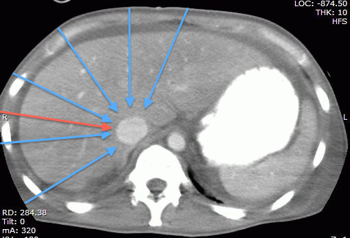
Technical Pearl: the part of the IVC we generally assess being the intrahepatic segment, it is possible to find it almost by scanning through any part of the liver, which happens to provide a great acoustic window. This may be particularly useful when the epigastric area is difficult to access (incision/bandage, drains, in the OR, etc…) or when there is bowel gas in the epigastrium. The figures below show the same IVC, first in a “traditional” epigastric view, then in a view approximately along the plane of the red arrow on the CT scan.

2 views IVC std:liver

CT liver IVC views


Bottom line?

When assessing volume status, it is absolutely essential to keep the clinical question in mind. It is a great minority of patients who are volume responsive who actually need volume. Normal, healthy humans are very much fluid responsive and fluid tolerant but certainly not in need of any. Much of the current studies and literature focus on assessing volume responsiveness in the setting of shock, which, although arguably the most important, is not the only type of information that can be obtained from the IVC. For instance, as will be discussed in the chapter on congestive heart failure, knowing that your patient is very “full” should prompt further diuresis. If you are dealing with managing severe anasarca, knowing that a patient’s intravascular volume is low may prompt the use of albumin or hypertonics prior to further diuresis to help resorb some of the interstitial fluid. Another critical question to which there is currently no answer is just how much fluid to give to patients in shock. There are many opinions but no certainty. It is common practice to fill a patient in shock until they are no longer fluid responsive, in an effort to avoid or minimize the use or dose of vasopressor medications. There is no study to date that compares a “moderate fluid/early vasopressors” vs “aggressive fluid/avoid-vasopressors-if-possible” approach.

Hopefully this will be answered soon. In light of the clear evidence linking positive fluid balance and mortality, it would seem wise to fill to a “moderate fullness” where some respiratory variation remains, rather than to the point of no longer being fluid responsive. After all, physiologically, the only time humans are really full (>20mm IVC with little or no variation) is in pathological states of congestive heart failure or obstructive shock. So again, when assessing an IVC, keep in mind what your clinical question is and interpret the sonographic data accordingly.


cheers!


Comments:

Marco says:

Thanks Philippe, very interesting post.
Using your great categorization, I think that the worst scenario is represented by a patient who is volume responsive but poorly volume tolerant. In that case, it is important to have clear in mind which is our target: cerebral perfusion, oxygenation and lung extravascular fluid, ventilatory weaning, renal function, avoiding vasopressors or mechanical ventilation, etc…
Very often, maintaining a brain dead heart beating donor, every organ would require a different volemic status, and if all of them are suitable for transplantation you have to compromise.
Just a last thought: as well as “all sniffs are not created equal”, also tidal volumes are very variable. Assessing respiratory variations during protective (low TV) ventilation requires a thoughtful interpretation of the results.


Absolutely. Interpretation of the findings in each individual case is key. 

Philippe

NEJM: The Septic Shock Issue…groundbreaking or same old same old? #FOAMed, #FOAMcc

Ok, so it was pretty cool to see an NEJM issue basically dedicated to septic shock management, I must admit. But let’s dig a little deeper, shall we?

So here is where they are: http://www.nejm.org, and fully available for now.

I won’t go through all the details and numbers, after all they are in the papers, so let’s just analyze them from two principles:

a. the N=1 principle – how was therapy individualized?

and

b. was there any integrated monitoring of the therapeutic goals?

…and we’ll conclude by looking at the potential practice-changing potential of each of these studies.

So first of all,

High vs Low BP Target in Septic Shock, by Asfar et al.

So basically a negative study except for two findings, the increased incidence of afib in the high target group and the decreased need for renal replacement therapy among chronic hypertensives in the high target group.

so N=1 is not really revealed:

“Refractoriness to fluid resuscitation was defined as a lack of response to the administration of 30 ml of normal saline per kilogram of body weight or of colloids or was determined according to a clinician’s assessment of inadequate hemodynamic results on the basis of values obtained during right-heart catheterization, pulse-pressure measurement, stroke-volume measurement, or echocardiography (although study investigators did not record the values for these variables).”

So lets just hope that the variability evens itself out between the groups, since we don’t really know. The numbers don’t really tell the tale, because the average fluids received (10 liters over 5 days) could mean one patient got 15 and one got 5 – although let’s trust they followed the French Fluid Resus protocol…

So the atrial fibrillation makes total sense – more B agonism should result in that, and the decreased renal failure also does.

As the authors note, the actual BP averages were higher than planned. For those of us practicing critical care, we know most nurses titrating prefer having a little bit of extra BP – even when I prescribe MAP 65, I usually see the 70 or so unless I make a point to tell them. Understandable. They also note the underpowered-ness of their own study, but I think it is still worth looking at their results.

So…bottom line?  I think it’s a great study for a couple of reasons.

The first is to remind us to pay a little more N=1 attention to the chronic hypertensives, and that it is probably worth aiming for slightly higher MAPs.

The second, debunking the myth of “levophed, leave’em dead” (which I heard throughout residency at McGill), and the concept of doing everything (ie juicing patient into a michelin man) in order to avoid the “dreaded and dangerous” vasopressors. So really I think an alternative way to conclude this study is that it isn’t harmful to have higher doses of vasopressors. I think this is actually a really good study on which to base assessment of more aggressive vasopressor support vs fluid resuscitation, in the right patients.

It would have been interesting to have echo data on those who developed a fib – were they patients who had normal to hyperdynamic LVs who in truth did not need B agonism at all and would have been fine with phenylephrine?  Perhaps…

Cool. I like it.

Next:

Albumin Replacement in Patients with Severe Sepsis or Septic Shock, by Caironi et al. The ALBIOS study (a Gattinoni crew)

So basically showed no difference, so pretty much a solid italian remake of the SAFE study in a sense, confirming that albumin is indeed safe overall, and may be better in those with shock.  As the authors note, mortality was low, organ failure was low, so study power a little low as well. Note the mean lactates in the 2’s at baseline. The albumin levels of the crytalloid only gorup were also not that low, low to mid 20’s, whereas I often see 15-20 range in my patients, especially if I inherit them after a few days, as I do use albumin myself a fair bit. They also used a target albumin level, not albumin as a resuscitation fluid purely.

In my mind the benefit of albumin would be greatest in those with significant capillary leak, particularly those with intra-abdominal and pulmonary pathology. It would have been nice to see a subgroup analysis where extravascular lung water was looked at (especially coming from a Gattinoni crew!).

Another interesting thing would have been to know the infusion time of the albumin, since animal data tells us that a 3hr infusion decreases extravasation and improves vascular filling vs shorter infusion times. I routinely insist on 3hr infusion per unit, which sometimes results in 9-12hr infusions, almost albumin drips!

Bottom line?

I like it. Reinforces that albumin is safe, so makes me even more comfortable in using it in the patients where my N=1 analysis tells me to be wary of third-spacing. Also the fact that they used 20% – in Canada we have 100cc bottles of 25% for the most part – is nice, since the SAFE data used 4%.

Next!

A Randomized Trial of Protocol-Based Care for Early Septic Shock – The ProCESS Trial.

So right off that bat my allergy to protocols flares up, so I’ll try to remain impartial. It just goes against the N=1 principle. The absolutely awesome thing about protocols is that it primes the team/system to react – so clearly protocols are better than no-protocol-at-all, but strict adherence would clearly not fit everyone, so that some built-in flexibility should be present.

This being said, the ProCESS study is really interesting, for a number of reasons. They have three groups, and compare basically (1) Rivers’ EGDT to (2) their own protocol (see the S2 appendix online) which gives a little more flexibility and (3) “usual care”.  Net result is that all are pretty equal, no change in mortality. As the authors note, their mortality was low, so again may not have been able to detect a difference.

So, what does this mean. To me it’s a little worrisome because I doubt that the “usual care” represents the true usual care found in EDs/ICUs all over the world, so I am concerned that many docs will use this as a reason to justify not changing their practice, similarly to many I’ve heard say they don’t need to cool anymore after the TTM trial. Human nature for some I guess.

Bottom line? You don’t have to follow EGDT if you’re conscientious and reassessing your patient frequently and have done all the other good things (abx, source control, etc). I think that’s really important because giving blood (see my post about S1P) to those with hb > 70 and giving dobutamine to patients with potentially normal or hyper dynamic LVs never made physiological sense to me, and the problem with a multi intervention study such as EGDT is that you can’t tease out the good from the bad or the neutral. Again, studies such as EGDT are pivotal in changing practice and raising awareness, so this is not a knock against a necessary study, just to highlight the point that each study is a step along the way of refining our resuscitation, and the important thing is to move on. In fact, the reason that this is a negative study is probably due to the improvement in “usual care” that EGDT brought along.

Conclusion: No new ground broken, but these studies do make me feel more confident and validated in continuing to not do certain things (strict EGDT) and  doing others (albumin and earlier use of vasopressors).

Kudos to all investigators.


let me know what you think!


P


Central line insertion: US-spotted “Blind” technique Video (HERE IT IS!) #FOAMed, #FOAMcc

My apologies, had technical issues with the video uploading so here it is in two formats:


…please let me know if there is a problem!


thanks


Philippe

CCUS 2014: Taking your Ultrasound Game to another level! Montreal may 9-11, 2014!

Registration is open!

For the 7th edition of our Annual Symposium, we’ve assembled a great cast of characters to bring your bedside ultrasound game to a whole new level! Whether you’re a novice or have some experience in bedside ultrasound, we’re sure you’ll find our program very interesting, as the perspective we have chosen to take is patient-based, with each lecture explaining how ultrasound findings are integrated into clinical decision-making.

First of all, it’s in Montreal, May 10th and 11th, 2014. If you’re from out of town, its an awesome city to visit, and if you’re a Montrealer, well, it’s an awesome conference at home!

Indeed, this isn’t “just” a “how-to” course – although that is what we’ll be covering in the workshops which will follow each lecture – but a “how to integrate ultrasound into your daily practice” course, so no matter what your level is, you’ll find something suited for you. The workshops will be divided into novice and experienced and with a low participant to faculty ratio, will ensure a great experience.

So what are we going to be talking about?

In the CC/ED/hospital scene:

–       the patient with chest pain, by Max Meineiri

–       the patient with CHF, by Mike Stone

–       the patient with renal failure, by Sara Sebbag

–       the patient with a swollenleg, by Michael Woo

–       what’s my patient’s volume status? By Ashraf Fayad

–       the patient with a decreased level of consciousness, by Robert Chen

–       the patient in shock, by JF Lanctot and Max Valois

–       the patient with abdominal pain, by Haney Mallemat

–       the patient with a bowel obstruction

–       the patient with broken bones: finding and freezing, by Catherine Nix

–       the patient with dyspnea, by Alberto Goffi and Edgar Hockmann (KEYNOTE)

–       the hemodynamically unstable patient: multi-modality assessment, by Andre Denault (KEYNOTE)

In the office scene:

–       the patient with a neck mass, by Sarah Sebbag

–       the patient with arthritis, by Alessandra Bruns

Here is the program:

CCUS 2014 Master Programme

Who made this up?  Our scientific committee is made up of Michael Woo, Catherine Nix, Edgar Hockmann, Alberto Goffi and Andre Denault, who is also the co-chair – with myself – of this year’s symposium.

For those of you part of the twitterverse and #FOAMed movement, you’ll get a chance to meet @criticalcarenow (Haney), @bedsidesono (Mike), @nobleultrasound (Vicki), @EGLS_JFandMax (JF and Max) and @ThinkingCC (yours truly)…and Matt (pulmccm.org) will be there to chronicle the event and bring the highlights to the #FOAMed world.

So who should come?  Anyone taking care of sick patients.  If you’re an ED doc, ICU doc, hospitalist, anaesthetist, surgeon, student or resident, this stuff is gold. Even if most of your practice is office-based, precise assessment of dyspnea, volume status and cardiac function is highly practical.

Additionally, there are several very interesting pre-congress courses taking place on May 9th, particularly the Echo-Guided Life Support course (JF Lanctot and Max Valois), a Bedside Ultrasound Course for Nurses, an Acute Care Procedures Course and a Symposium for Ultrasound Educators on how to set up training programs. All in all, a lot of great stuff.

For more details and registration, please go to www.ccusinstitute.org.  And yes, of course there are CME credits by the University of Montreal.

As I said a few months ago – well, tweeted, to be exact – if you’re an acute care MD and NOT using bedside point-of-care ultrasound, you’re stuck in the 20th century… rude, but true.

Hope to see you there!

Philippe Rola

President, CCUS Institute

Pleural effusion in the sick patient (Part 1of 3): Don’t miss it!!! #FOAMed, #FOAMcc

This, in my opinion, is an under-recognized problem when bedside ultrasound is NOT a routine part of examination of critically ill patients. I’m happy to say that as many of my colleagues have been picking up probes, it is somewhat less of an issue now, whereas a couple of years ago I’d often put in 4 or 5 pigtail catheters on day 1 of taking over the ICU.

The first and foremost reason for this is that the portable supine ICU CXR sucks at picking up the small to moderate to, yes, even the large pleural effusion.  Largely owing to the fact that many of our patients have some lung parenchymal abnormalities and to the recumbent position that causes a layering of the effusion, it is often difficult to properly assess the size of a pleural effusion.  Radiologists will usually report the presence of a probable effusion, but quantification is difficult, and physicians not performing routing bedside sonography will often realize the presence of a submassive effusion only on CT scan – after all it isn’t like you can turn and rotate your patient to percuss the shifting dullness, can you?  Not very practical.

So the following can often be seen:

pleural effusion

this is fairly large, or you might see:

pleural effusion and pneumonia

So the obvious and critical question is: when is it necessary to drain?

There are two elements to this question:

a) for diagnostic purposes: unless the diagnosis is clear (eg CHF, post-resuscitation “michelin man” patient, etc) a new effusion should be tapped.  Panapneumonic effusions, in particular, warrant ruling out empyema unless there is a compelling reason not to.  For diagnostic purposes a 22g needle usually does the trick unless you have frank pus – which generally shows up differently on ultrasound.

b) for therapeutic purposes: effusions are space occupying lesions which compress the lung and result in a variable degree of respiratory compromise, depending on chest wall and diaphragmatic compliance, as well as effusion volume.  The clinical effect is highly variable due to the above as well as the degree of parenchymal lung disease and the degree of PEEP.  In the ICU or ED, a simple way to think about it is that if your patient is in respiratory failure and has a large effusion, chances are that draining it will improve things.  It gets a little more controversial and complicated if you have a patient who is mildly dyspneic with a moderate sized effusion.

Here are a few clinical scenarios I like:

Mr. A is a 65 year old man with CHF, intubated, with large bilateral effusions. He has been aggressively diuresed to the point of his IVC being less than 5mm in diameter.  He has not been able to wean in the last 48 h.

Yes, I definitely drain this fellow. Been there and done that time and time again.  The pleural effusions are essentially the last to resolve (being the most “distal” to the circulation – vs the alveolar tissue itself) and hence can lag and cost a few more days or more of ventilation).

Mr. B is a 47 year old man with pneumonia, breathing spontaneously with a moderate (maybe 500ml) effusion. It appears free flowing and clear, he is afebrile with an improving white count, and mildly dyspneic.

Nah, I skip on this one.  If fever and WBC recur, I do a diagnostic tap to r/o empyema.

Mrs. C is intubated on PEEP 18 FiO2 85% for ARDS due to pancreatitis. She has some degree of intraabdominal hypertension (IAP 18) and has bilateral moderate pleural effusions, maybe 400-500ml.

Yup. She can physiologically benefit from decreased intra-thoracic pressure, both from the ventilatory and the intra-abdominal pressure standpoint (Remember the diaphragm is not a rigid structure so that IAP and ITP are very similar in most cases).

So is there any evidence for this?  Some. And that’s for part 2, coming within the next days. Part three will explain and show my procedure of choice for drainage.

Thanks!

love to hear what other guys’ practices are!   Apparently only about 15% of ICU guys “routinely” drain effusions.

Philippe

COMMENTS

Hi Philippe,
I am very happy to read your post tonight, bacause I am part of that 15% and luckily most of my colleagues are in the same group. I agree in particular when you say that pleural effusions are the last to resolve, being the most “distal” to the circulation. I often find patients, at a certain point during their ICU stay, be not only like a “michelin man” but also (and at the same time!) hypovolemic. I call this situation, when I try to explain it to residents, “empty in full” (maybe in english it doesn’t sound as good as in italian): we are trying with diuretics and some fluid restriction to manage those extravascular fluids that prevent weaning from mechanical ventilation, but often we get the only effect of causing renal failure rather than eliminating pleural effusions. In this case the only way is to drain.
Another important point is that bedside chest x-ray is absolutely useless when you have to discriminate between pleural effusion and parenchimal consolidation, both of them often coexisting in ICU patients.
In our routine we use 14-gauge single lumen CVCs, inserted with Seldinger technique and ultrasound assisted procedure, effective in 95% of the effusions and less invasive than a pleurocath (that we use most of the times for pneumothorax) or a real chest tube, which I keep for blood or traumatic pneumothorax.
Greetings from Italy,
Marco


Glad to hear it Marco!  I started with CVCs as well before we were able to find inexpensive pigtails – email me for info if you want I don’t want to “brand” these things!  They are actually really good because I found CVCs would often occlude. Patient comfort with either is so much more than chest tubes. 

Great point about the “michelin man” who is very “wet” but intravascularly dry, which we see commonly post acute phase of critical illness, especially when physicians are so keen to use crystalloids.

thanks for reading!


Philippe

Blood transfusion and serum S1P levels in Sepsis: a leaky proposition? (Protecting the Glycocalyx Part1) #FOAMed, #FOAMcc

So in my ongoing quest to reframe my resuscitation step-by-step, I’ve been following up on a number of leads regarding the glycocalyx, as previously stated, and John’s reference to this article in a previous comment I feel is highly relevant. So this is it:

Synergistic Effect of Anemia and Red Blood Cells Transfusion on Inflammation and Lung Injury,  Anping Dong, Manjula Sunkara, Manikandan Panchatcharam, Abdel Salous, Samy Selim, Andrew J.Morris, and Susan S. Smyth

Advances in Hematology, Volume 2012, Article ID 924042, 8 pages

http://www.hindawi.com/journals/ah/2012/924042/

S1P (sphingosine-1-phosphate) is a regulator of endothelial permeability and immune function.  Uh-oh, why had I not heard of it? Hmmm…I don’t think it was in Guyton’s or in Harrison’s…and there hasn’t been an RCT about it… Ok, that about explains it.

So here are some factoids about S1P:

–       serum levels correlate with HCT as RBCs serve as an S1P reservoir.

–       anemic patients’ S1P levels are NOT fully replenished by transfusion, especially the older the transfused blood is.

–       In fact, older RBCs may actually remove plasma S1P.

The study:

They basically took mice, and in the first group, bled them (by 20ml/kg) and looked at inflammatory markers, lung permeability and also S1P levels. That’s basically the control group, and they noted that hemorrhage significantly increased inflammatory markers (interesting in and of itself) . They then transfused these mice using wither fresh, S1P-loaded RBCs, or 14-day old RBCs, and, lo and behold, the fresh blood resulted in less inflammation, increased S1P, but most importantly, markedly decreased lung permeability. So clearly, S1P attenuates transfusion associated lung permeability.

In the next group, they injected the mice with LPS following hemorrhage, and found a synergistic effect of blood loss and LPS on inflammation and lung permeability, as could be imagined. Following the LPS, they were transfused with one of four strategies: fresh blood, old blood, fresh blood + S1P or old blood + S1P.  Well, lung permeability still increased in all groups, but least in the fresh blood + S1P, and the old blood + S1P a close second.

Note, interestingly enough, that saline alone (the “control”) also increased lung permeability, highlighting yet again that NS (and probably any crystalloid) is not innocuous…

So here we’re looking at the finer effects of transfusion, and why, against “common-sense” correcting a patient’s hemoglobin level does not seem to help in all situations.  We have understood the aging issue and loss of deformability, but it is time to take a finer look.  We are familiar – at least in concept – with transfusion-associated lung injury or TRALI, but the mechanism remains unclear.

Summary and Take-Home message:

S1P infusions in sepsis?  Maybe someday…

Yes, this is an animal study, and the results cannot be extrapolated directly to humans, but it is food for thought, as John had mentioned.  Certainly at least this should tell us to keep and ear/eye out for human work with S1P, but personally, it will make me even more comfortable in not transfusing my septic patients with hb’s in the low 70’s and maybe even high 60’s (try repeating the cbc, more often than not comes back a couple points higher and you can avoid transfusion), and for those who are a little more aggressive with transfusion, maybe this should make them think twice…

I’ll add what I can dig up on human S1P studies soon.

cheers!

Philippe

Bedside Ultrasound Clip Quiz #7: What is it? #FOAMed, #FOAMcc

Ok, so no history for you. Say you’re doing a general bedside ultrasound exam on someone in shock and you see this:

What am I looking at?  (HINT we’re in the RUQ)

What should I be thinking of?

Answers tomorrow.  Lets see what you guys come up with!

Philippe

rubbadoc says:  What’s air doing there?

…and indeed he is right, it is air where there should not be. Now where is it exactly? (experts, refrain from answering…)